Copyright
copy;2010 Baishideng Publishing Group Co.
World J Gastroenterol. Sep 28, 2010; 16(36): 4594-4598
Published online Sep 28, 2010. doi: 10.3748/wjg.v16.i36.4594
Published online Sep 28, 2010. doi: 10.3748/wjg.v16.i36.4594
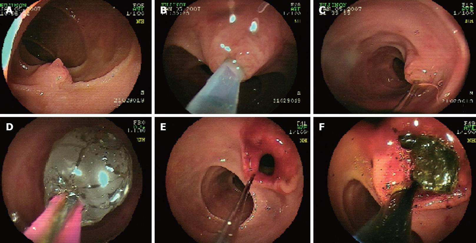
Figure 2 Endoscopy showing the papilla reached (A) and successfully cannulated (B) by double balloon endoscope, the guide wire left in the bile duct (C), endoscopic papillary balloon dilation performed (D, E), and stones found using the balloon (F).
- Citation: Lin CH, Tang JH, Cheng CL, Tsou YK, Cheng HT, Lee MH, Sung KF, Lee CS, Liu NJ. Double balloon endoscopy increases the ERCP success rate in patients with a history of Billroth II gastrectomy. World J Gastroenterol 2010; 16(36): 4594-4598
- URL: https://www.wjgnet.com/1007-9327/full/v16/i36/4594.htm
- DOI: https://dx.doi.org/10.3748/wjg.v16.i36.4594


