Copyright
copy;2010 Baishideng Publishing Group Co.
World J Gastroenterol. Sep 28, 2010; 16(36): 4583-4588
Published online Sep 28, 2010. doi: 10.3748/wjg.v16.i36.4583
Published online Sep 28, 2010. doi: 10.3748/wjg.v16.i36.4583
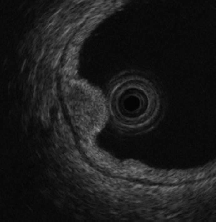
Figure 2 Upper gastrointestinal endoscopy showed a homogenous, oval hypoechoic mass, mainly located in the third layer.
- Citation: Ishido K, Tanabe S, Higuchi K, Sasaki T, Katada C, Azuma M, Naruke A, Koizumi W, Mikami T. Clinicopathological evaluation of duodenal well-differentiated endocrine tumors. World J Gastroenterol 2010; 16(36): 4583-4588
- URL: https://www.wjgnet.com/1007-9327/full/v16/i36/4583.htm
- DOI: https://dx.doi.org/10.3748/wjg.v16.i36.4583


