Copyright
copy;2010 Baishideng Publishing Group Co.
World J Gastroenterol. Sep 28, 2010; 16(36): 4549-4557
Published online Sep 28, 2010. doi: 10.3748/wjg.v16.i36.4549
Published online Sep 28, 2010. doi: 10.3748/wjg.v16.i36.4549
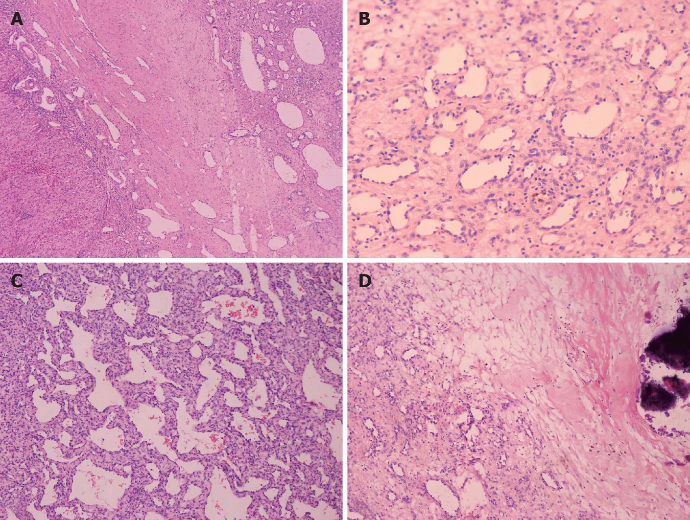
Figure 3 Histopathological features of type I lesions.
A: Mature vascular channels (case 7, HE, × 60); B: Plump endothelial cells with a bland cytological appearance (case 7, HE, × 200); C: Cavernous structures (case 7, HE, × 100); D: Calcification foci (case 7, HE, × 100).
- Citation: Zhang Z, Chen HJ, Yang WJ, Bu H, Wei B, Long XY, Fu J, Zhang R, Ni YB, Zhang HY. Infantile hepatic hemangioendothelioma: A clinicopathologic study in a Chinese population. World J Gastroenterol 2010; 16(36): 4549-4557
- URL: https://www.wjgnet.com/1007-9327/full/v16/i36/4549.htm
- DOI: https://dx.doi.org/10.3748/wjg.v16.i36.4549


