Copyright
copy;2010 Baishideng Publishing Group Co.
World J Gastroenterol. Sep 28, 2010; 16(36): 4549-4557
Published online Sep 28, 2010. doi: 10.3748/wjg.v16.i36.4549
Published online Sep 28, 2010. doi: 10.3748/wjg.v16.i36.4549
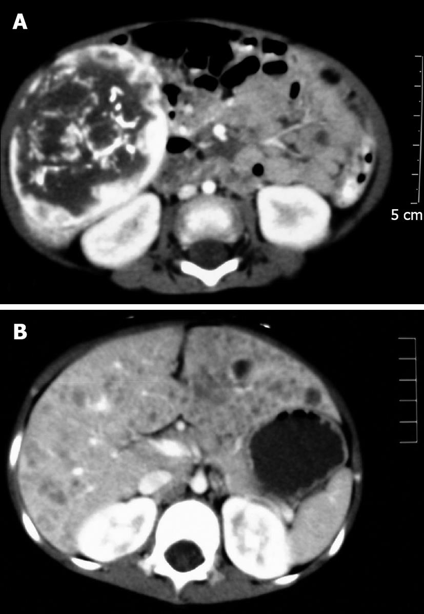
Figure 1 Contrast-enhanced arterial phase computed tomography.
A: Solitary hypoattenuated mass with a well-defined contour containing calcifications in a fine-speckled pattern (case 7); B: Diffuse polycystic changes with variable sizes and an ill-defined region in both hepatic lobes (case 5).
- Citation: Zhang Z, Chen HJ, Yang WJ, Bu H, Wei B, Long XY, Fu J, Zhang R, Ni YB, Zhang HY. Infantile hepatic hemangioendothelioma: A clinicopathologic study in a Chinese population. World J Gastroenterol 2010; 16(36): 4549-4557
- URL: https://www.wjgnet.com/1007-9327/full/v16/i36/4549.htm
- DOI: https://dx.doi.org/10.3748/wjg.v16.i36.4549


