Copyright
©2010 Baishideng Publishing Group Co.
World J Gastroenterol. Sep 14, 2010; 16(34): 4281-4290
Published online Sep 14, 2010. doi: 10.3748/wjg.v16.i34.4281
Published online Sep 14, 2010. doi: 10.3748/wjg.v16.i34.4281
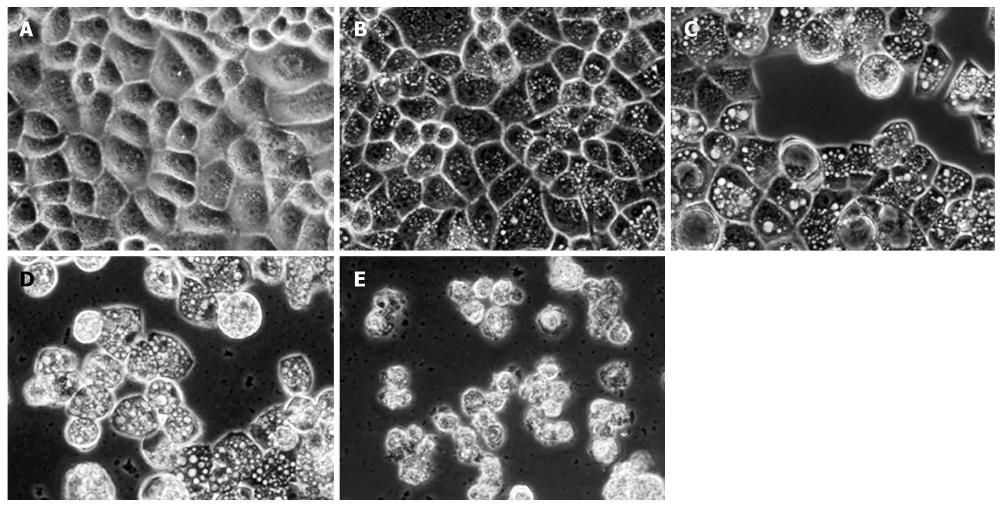
Figure 5 Inverted phase contrast microscopy showing matrine-induced morphologic changes of HepG2 cells.
The control cells were well adhered, displaying the normal morphology of HepG2 cells. In contrast, abundant cytoplasmic vacuoles were observed in cells treated with matrine. Moreover, vacuolization in cytoplasm progressively became larger and denser when the concentration of matrine was increased. The majority of HepG2 cells treated with matrine at the concentration of 2.0 mg/mL became round and shrunken and could not be affixed to the wall and suspended in culture medium (× 400 magnification). A: 0 mg/mL matrine; B: 0.25 mg/mL matrine; C: 0.5 mg/mL matrine; D: 1.0 mg/mL matrine; E: 2.0 mg/mL matrine.
- Citation: Zhang JQ, Li YM, Liu T, He WT, Chen YT, Chen XH, Li X, Zhou WC, Yi JF, Ren ZJ. Antitumor effect of matrine in human hepatoma G2 cells by inducing apoptosis and autophagy. World J Gastroenterol 2010; 16(34): 4281-4290
- URL: https://www.wjgnet.com/1007-9327/full/v16/i34/4281.htm
- DOI: https://dx.doi.org/10.3748/wjg.v16.i34.4281


