Copyright
copy;2010 Baishideng Publishing Group Co.
World J Gastroenterol. Sep 7, 2010; 16(33): 4237-4242
Published online Sep 7, 2010. doi: 10.3748/wjg.v16.i33.4237
Published online Sep 7, 2010. doi: 10.3748/wjg.v16.i33.4237
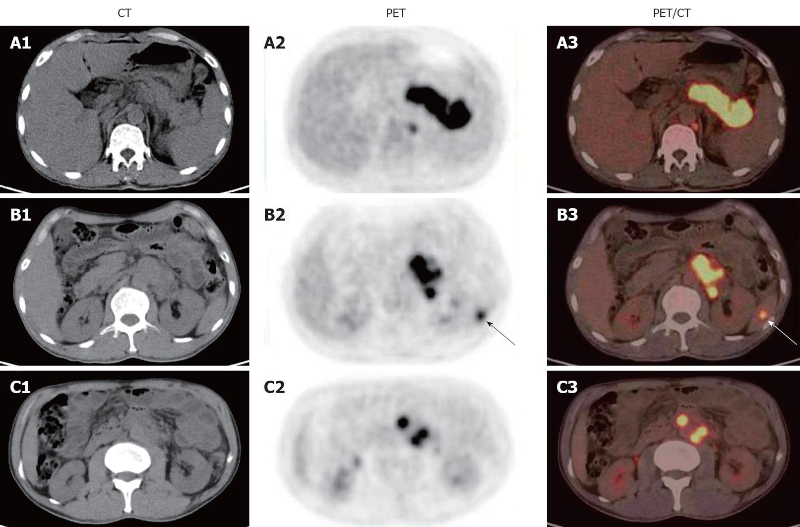
Figure 1 18F-fluorodeoxyglucose positron emission/computed tomography images of patient 1 showing a mass-like area with an intense 18F-fluorodeoxyglucose uptake extending up behind the pancreatic body and tail (A1-A3), a focal area with an intense 18F-fluorodeoxyglucose uptake in spleen (arrows) (B1-B3), and multiple focal areas with an intense 18F-fluorodeoxyglucose uptake around the abdominal aorta (C1-C3).
CT: Computed tomography; PET: Positron emission tomography.
- Citation: Tian G, Xiao Y, Chen B, Guan H, Deng QY. Multi-site abdominal tuberculosis mimics malignancy on 18F-FDG PET/CT: Report of three cases. World J Gastroenterol 2010; 16(33): 4237-4242
- URL: https://www.wjgnet.com/1007-9327/full/v16/i33/4237.htm
- DOI: https://dx.doi.org/10.3748/wjg.v16.i33.4237


