Copyright
copy;2010 Baishideng Publishing Group Co.
World J Gastroenterol. Sep 7, 2010; 16(33): 4145-4151
Published online Sep 7, 2010. doi: 10.3748/wjg.v16.i33.4145
Published online Sep 7, 2010. doi: 10.3748/wjg.v16.i33.4145
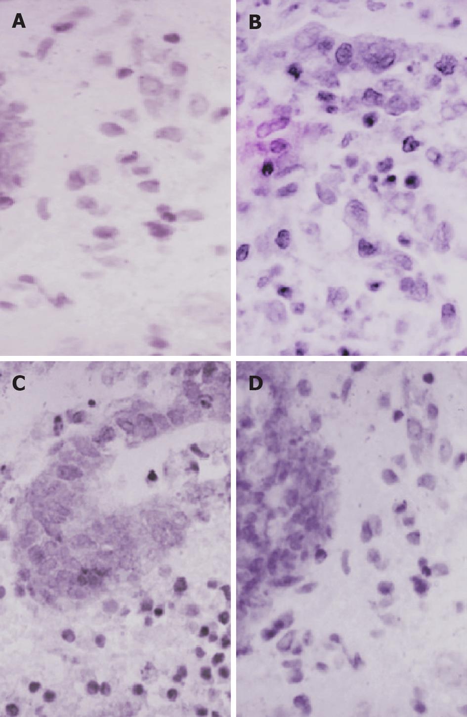
Figure 3 Immunohistochemical staining for nuclear factor-κB p65 (Brown staining, SP × 700).
A: Section of colon from controls showing normal structure and architecture; B: Section of colon from ulcerative colitis patients before treatment showing extensive nuclear factor (NF)-κB (brown) expression; C: Section of colon from sulfasalazine group showing limited NF-κB (brown) expression; D: Section of colon from probiotic group showing minimal NF-κB (brown) expression.
- Citation: Hegazy SK, El-Bedewy MM. Effect of probiotics on pro-inflammatory cytokines and NF-κB activation in ulcerative colitis. World J Gastroenterol 2010; 16(33): 4145-4151
- URL: https://www.wjgnet.com/1007-9327/full/v16/i33/4145.htm
- DOI: https://dx.doi.org/10.3748/wjg.v16.i33.4145


