Copyright
©2010 Baishideng Publishing Group Co.
World J Gastroenterol. Aug 28, 2010; 16(32): 4107-4111
Published online Aug 28, 2010. doi: 10.3748/wjg.v16.i32.4107
Published online Aug 28, 2010. doi: 10.3748/wjg.v16.i32.4107
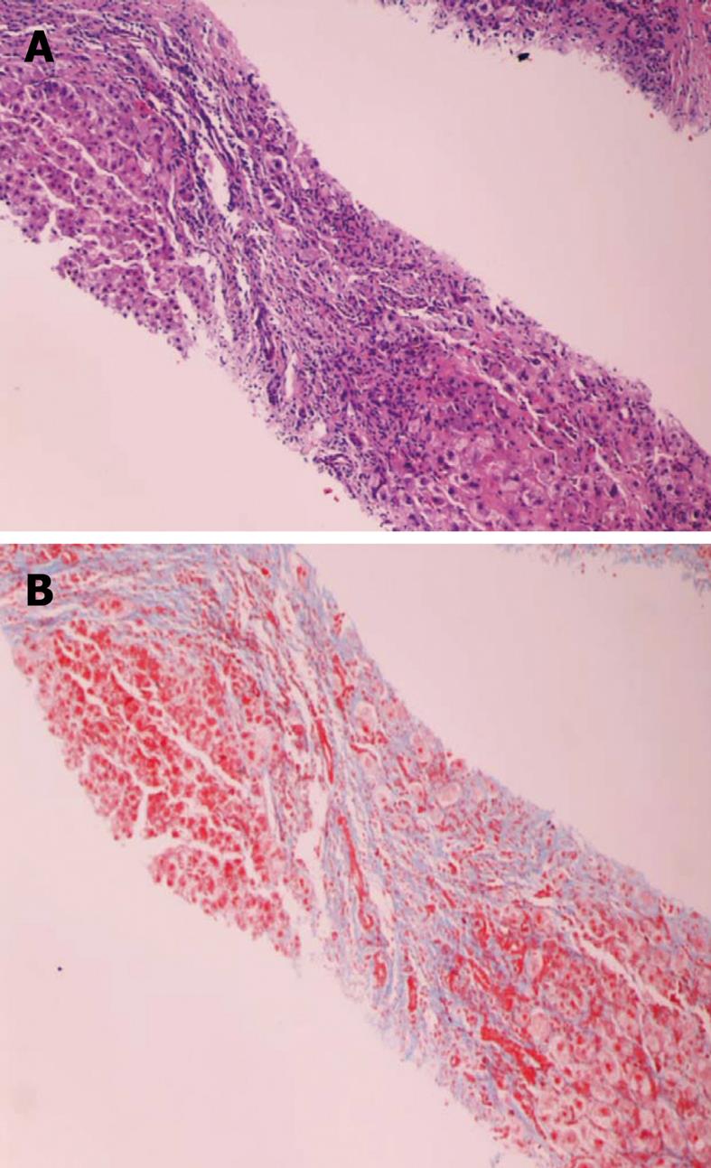
Figure 1 Liver biopsy, low power magnification (100 ×).
A: Hematoxylin and eosin stain showing a low power view of two cirrhotic nodule surrounded by fibrosis. The edges of the nodules and the fibrous septi contain intense chronic inflammatory infiltrate; B: Trichrome stain delineating fibrous septa that surround liver nodules in blue color.
- Citation: Anderson AM, Mosunjac MB, Palmore MP, Osborn MK, Muir AJ. Development of fatal acute liver failure in HIV-HBV coinfected patients. World J Gastroenterol 2010; 16(32): 4107-4111
- URL: https://www.wjgnet.com/1007-9327/full/v16/i32/4107.htm
- DOI: https://dx.doi.org/10.3748/wjg.v16.i32.4107


