Copyright
©2010 Baishideng.
World J Gastroenterol. Aug 21, 2010; 16(31): 3979-3983
Published online Aug 21, 2010. doi: 10.3748/wjg.v16.i31.3979
Published online Aug 21, 2010. doi: 10.3748/wjg.v16.i31.3979
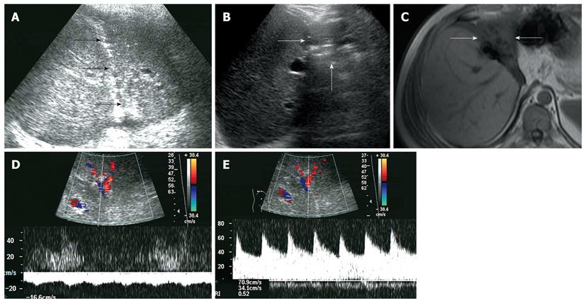
Figure 3 A 37-year-old man with a shranken left graft after dual-graft liver transplantation.
A: Gray scale sonogram shows the left graft (arrows) on postoperative day one; B and C: Size of the left graft decreased on follow-up sonogram and magnetic resonance imaging (arrows); D: Doppler spectrum shows the hepatofugal flow of the portal vein of left graft; E: Doppler spectrum shows the normal left hepatic artery
- Citation: Lu Q, Wu H, Yan LN, Chen ZY, Fan YT, Luo Y. Living donor liver transplantation using dual grafts: Ultrasonographic evaluation. World J Gastroenterol 2010; 16(31): 3979-3983
- URL: https://www.wjgnet.com/1007-9327/full/v16/i31/3979.htm
- DOI: https://dx.doi.org/10.3748/wjg.v16.i31.3979


