Copyright
©2010 Baishideng.
World J Gastroenterol. Aug 21, 2010; 16(31): 3964-3969
Published online Aug 21, 2010. doi: 10.3748/wjg.v16.i31.3964
Published online Aug 21, 2010. doi: 10.3748/wjg.v16.i31.3964
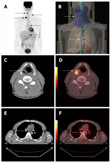
Figure 3 A 53-year-old woman with synchronous hypopharynx cancer combined with esophageal cancer.
Positron emission tomography/computed tomography (PET/CT) images revealed hypermetabolic hypopharynx and esophageal lesions, and multiple hypermetabolic supraclavicular, mediastinal lymph nodes (arrows in A). PET/CT imaging guided radiation plan (arrows in B). CT and PET/CT fused images showed hypermetabolic hypopharynx and esophageal lesions (arrows in C, D, E, F).
- Citation: Sun L, Wan Y, Lin Q, Sun YH, Zhao L, Luo ZM, Wu H. Multiple primary malignant tumors of upper gastrointestinal tract: A novel role of 18F-FDG PET/CT. World J Gastroenterol 2010; 16(31): 3964-3969
- URL: https://www.wjgnet.com/1007-9327/full/v16/i31/3964.htm
- DOI: https://dx.doi.org/10.3748/wjg.v16.i31.3964


