Copyright
©2010 Baishideng.
World J Gastroenterol. Aug 21, 2010; 16(31): 3928-3935
Published online Aug 21, 2010. doi: 10.3748/wjg.v16.i31.3928
Published online Aug 21, 2010. doi: 10.3748/wjg.v16.i31.3928
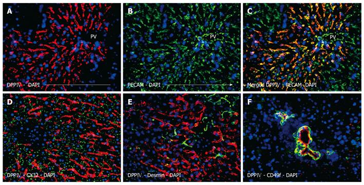
Figure 3 Phenotypic assessment of the non-hepatocytes liver repopulation showing extensive and string-like repopulation by liver sinusoidal endothelial cells emerging from the portal veins.
Donor derived cells were identified by dipeptidyl peptidase IV (DPPIV) immunofluorescence staining (red) (A), which co-localized with the endothelial marker PECAM (green) (B), overlay (C). Donor endothelial cells could be clearly distinguished from endogenous hepatocytes which were outlined by the hepatic differentiation marker CX32 (green) (D), desmin-positive cells (green) were detectable with a regular staining pattern for normal (unharmed) liver (E). Additionally, DPPIV-positive donor cells formed bile duct structures which expressed the specific marker CD49f (green) (C), nuclear counterstaining with DAPI (blue), original magnification × 200.
- Citation: Krause P, Rave-Fränk M, Wolff HA, Becker H, Christiansen H, Koenig S. Liver sinusoidal endothelial and biliary cell repopulation following irradiation and partial hepatectomy. World J Gastroenterol 2010; 16(31): 3928-3935
- URL: https://www.wjgnet.com/1007-9327/full/v16/i31/3928.htm
- DOI: https://dx.doi.org/10.3748/wjg.v16.i31.3928


