Copyright
©2010 Baishideng.
World J Gastroenterol. Aug 21, 2010; 16(31): 3928-3935
Published online Aug 21, 2010. doi: 10.3748/wjg.v16.i31.3928
Published online Aug 21, 2010. doi: 10.3748/wjg.v16.i31.3928
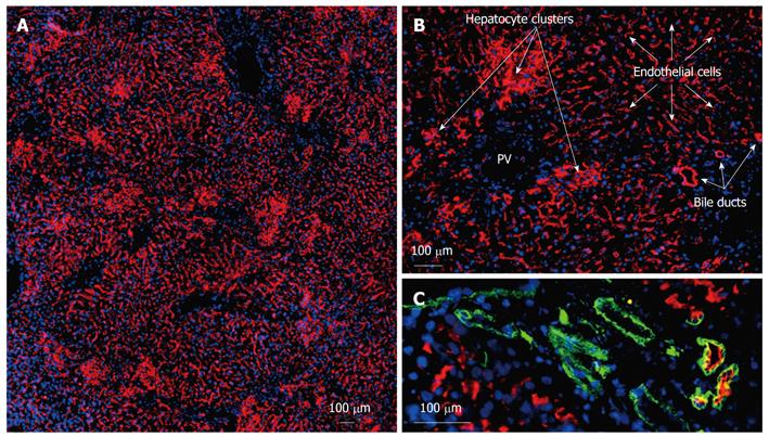
Figure 2 Characteristics of dipeptidyl peptidase IV-positive cells in the liver following transplantation of non-parenchymal cell preparations and hepatocytes.
Frozen sections were obtained from recipient livers present at 16 wk post-transplantation. Acetone-fixed frozen sections were single or double-labeled by indirect immunofluorescence analysis with an antibody detecting the donor cell specific dipeptidyl peptidase IV (red) (A-C) and bile-duct specific anti-CD49f (green) (C). Merged images are combined with blue nuclear DAPI-staining. Circumscribed and compact clusters of hepatocyte repopulation could be distinguished from large areas of endothelial repopulation which covered a maximum of 90% of the section plane. Formation of bile duct-like structures could be detected by co-staining with the duct marker CD49f. PV: Portal vein.
- Citation: Krause P, Rave-Fränk M, Wolff HA, Becker H, Christiansen H, Koenig S. Liver sinusoidal endothelial and biliary cell repopulation following irradiation and partial hepatectomy. World J Gastroenterol 2010; 16(31): 3928-3935
- URL: https://www.wjgnet.com/1007-9327/full/v16/i31/3928.htm
- DOI: https://dx.doi.org/10.3748/wjg.v16.i31.3928


