Copyright
©2010 Baishideng.
World J Gastroenterol. Aug 21, 2010; 16(31): 3928-3935
Published online Aug 21, 2010. doi: 10.3748/wjg.v16.i31.3928
Published online Aug 21, 2010. doi: 10.3748/wjg.v16.i31.3928
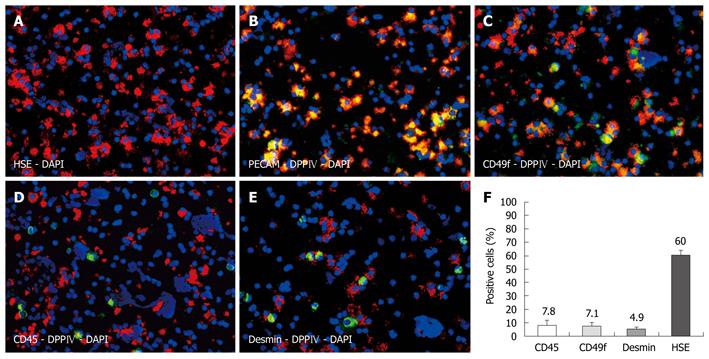
Figure 1 Characterization of non-parenchymal cell preparations by immunofluorescence on cytospins.
Hepatic sinusoidal endothelial (HSE) (red) identifying approximately 60% of all cells as hepatic sinusoidal cells (A), donor specific dipeptidyl peptidase IV (DPPIV) (red) co-localized with platelet endothelial cell adhesion molecule (green) (B), and with CD49f as bile duct marker (green) (C). Donor cells were negative for CD45 (D) and desmin (E). Labeling indices (F), nuclear counterstaining with DAPI (blue), original magnification × 200.
- Citation: Krause P, Rave-Fränk M, Wolff HA, Becker H, Christiansen H, Koenig S. Liver sinusoidal endothelial and biliary cell repopulation following irradiation and partial hepatectomy. World J Gastroenterol 2010; 16(31): 3928-3935
- URL: https://www.wjgnet.com/1007-9327/full/v16/i31/3928.htm
- DOI: https://dx.doi.org/10.3748/wjg.v16.i31.3928


