Copyright
©2010 Baishideng.
World J Gastroenterol. Aug 21, 2010; 16(31): 3911-3918
Published online Aug 21, 2010. doi: 10.3748/wjg.v16.i31.3911
Published online Aug 21, 2010. doi: 10.3748/wjg.v16.i31.3911
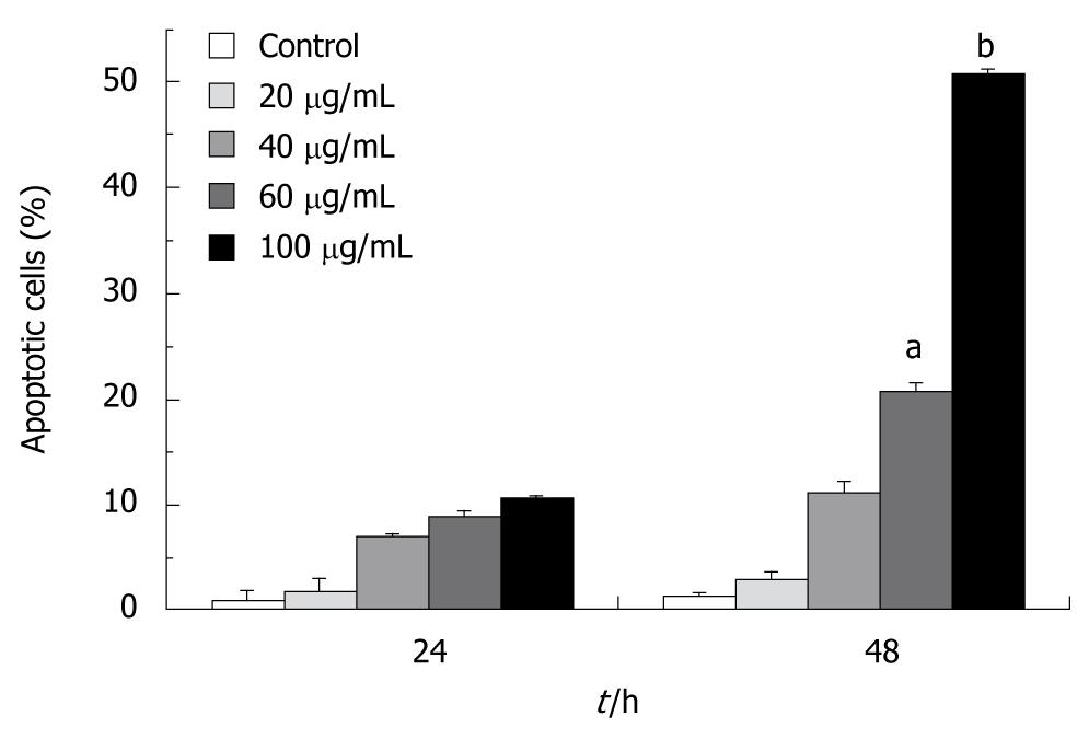
Figure 4 Flow cytometry-evidenced apoptosis of hepatic stellate cells-T6 cells upon exposure to tectorigenin.
Cells were incubated for 24 and 48 h with tectorigenin at 20, 40, 60 and 100 μg/mL, respectively, followed by being stained with fluorescein isothiocyanate-conjugated annexin V and propidium iodide. Bars represent mean ± SD (aP < 0.05, bP < 0.01).
- Citation: Wu JH, Wang YR, Huang WY, Tan RX. Anti-proliferative and pro-apoptotic effects of tectorigenin on hepatic stellate cells. World J Gastroenterol 2010; 16(31): 3911-3918
- URL: https://www.wjgnet.com/1007-9327/full/v16/i31/3911.htm
- DOI: https://dx.doi.org/10.3748/wjg.v16.i31.3911


