Copyright
©2010 Baishideng.
World J Gastroenterol. Aug 21, 2010; 16(31): 3911-3918
Published online Aug 21, 2010. doi: 10.3748/wjg.v16.i31.3911
Published online Aug 21, 2010. doi: 10.3748/wjg.v16.i31.3911
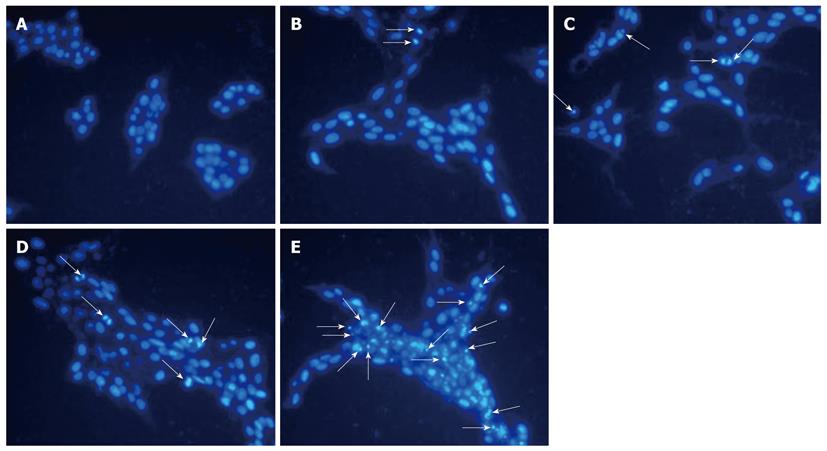
Figure 3 Fluorescent staining of nuclei in hepatic stellate cells-T6 cells (200 ×).
Cells were treated with tectorigenin for 48 h at 0 μg/mL (A), 20 μg/mL (B), 40 μg/mL (C), 60 μg/mL (D) and 100 μg/mL (E), respectively. The arrows in B-E indicate the apoptotic cells.
- Citation: Wu JH, Wang YR, Huang WY, Tan RX. Anti-proliferative and pro-apoptotic effects of tectorigenin on hepatic stellate cells. World J Gastroenterol 2010; 16(31): 3911-3918
- URL: https://www.wjgnet.com/1007-9327/full/v16/i31/3911.htm
- DOI: https://dx.doi.org/10.3748/wjg.v16.i31.3911


