Copyright
©2010 Baishideng.
World J Gastroenterol. Jan 21, 2010; 16(3): 398-402
Published online Jan 21, 2010. doi: 10.3748/wjg.v16.i3.398
Published online Jan 21, 2010. doi: 10.3748/wjg.v16.i3.398
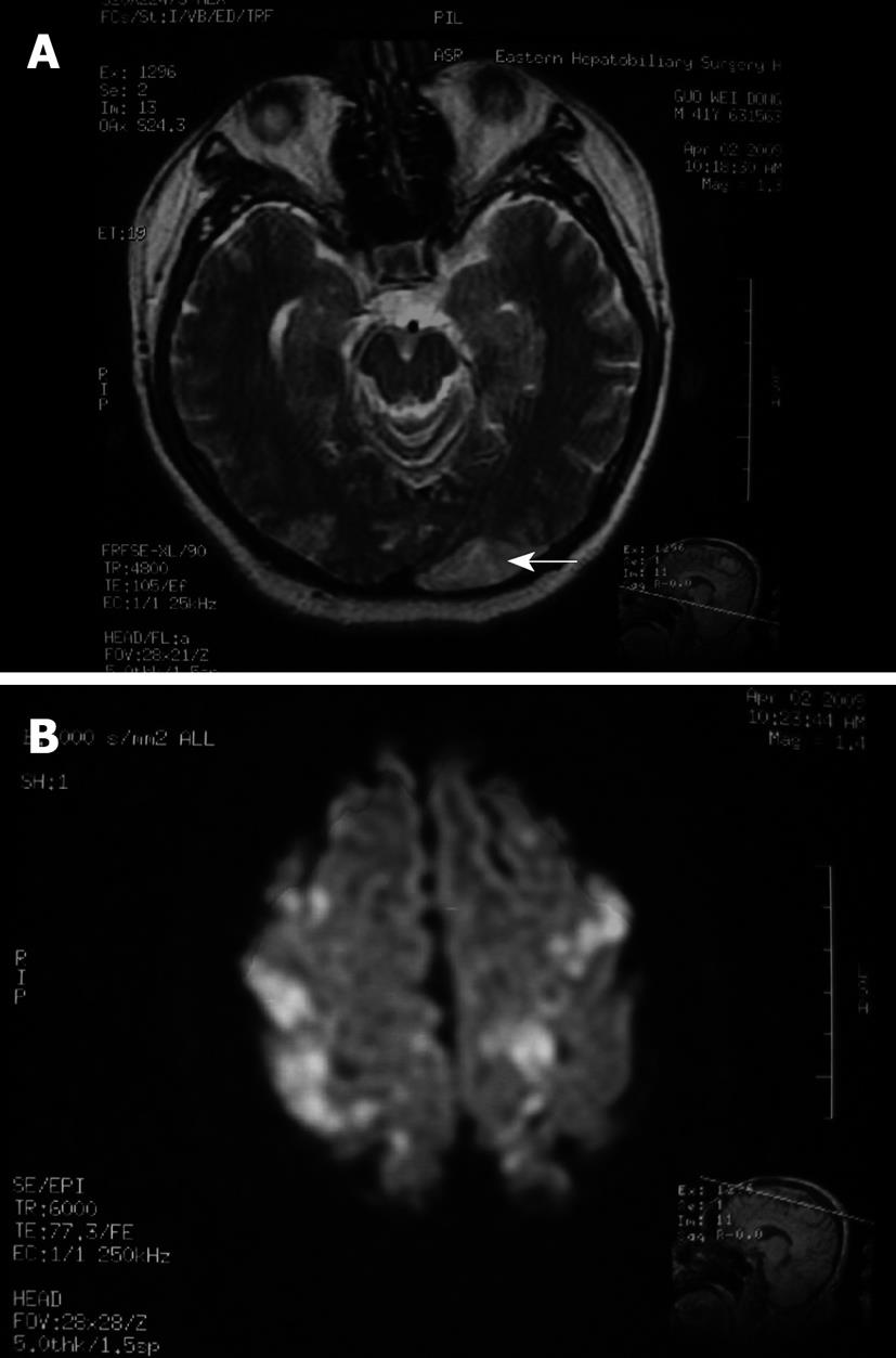
Figure 4 Cranial magnetic resonance imaging (MRI) obtained after the second TACE procedure.
A: cerebral lipiodol embolism (arrow); B: cerebral lipiodol embolism (multiple high signals).
- Citation: Wu L, Yang YF, Liang J, Shen SQ, Ge NJ, Wu MC. Cerebral lipiodol embolism following transcatheter arterial chemoembolization for hepatocellular carcinoma. World J Gastroenterol 2010; 16(3): 398-402
- URL: https://www.wjgnet.com/1007-9327/full/v16/i3/398.htm
- DOI: https://dx.doi.org/10.3748/wjg.v16.i3.398


