Copyright
©2010 Baishideng.
World J Gastroenterol. Aug 7, 2010; 16(29): 3727-3730
Published online Aug 7, 2010. doi: 10.3748/wjg.v16.i29.3727
Published online Aug 7, 2010. doi: 10.3748/wjg.v16.i29.3727
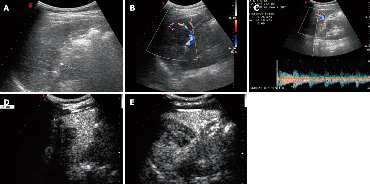
Figure 2 Conventional ultrasonography and contrast-enhanced ultrasonography findings of sclerosing angiomatoid nodular transformation in a 37-year-old woman with pain in the left upper quadrant.
A-C: A heterogeneous hypoechoic lesion (arrow) in the spleen, with blood-flow signals in the peripheral area; D: There was a 0.9 cm homologous nodule attaching to the side of the lesion that became evident after being enhanced (2 min 48 s after injection); E: The lesion was enhanced homogeneously.
- Citation: Cao JY, Zhang H, Wang WP. Ultrasonography of sclerosing angiomatoid nodular transformation in the spleen. World J Gastroenterol 2010; 16(29): 3727-3730
- URL: https://www.wjgnet.com/1007-9327/full/v16/i29/3727.htm
- DOI: https://dx.doi.org/10.3748/wjg.v16.i29.3727


