Copyright
©2010 Baishideng.
World J Gastroenterol. Aug 7, 2010; 16(29): 3664-3673
Published online Aug 7, 2010. doi: 10.3748/wjg.v16.i29.3664
Published online Aug 7, 2010. doi: 10.3748/wjg.v16.i29.3664
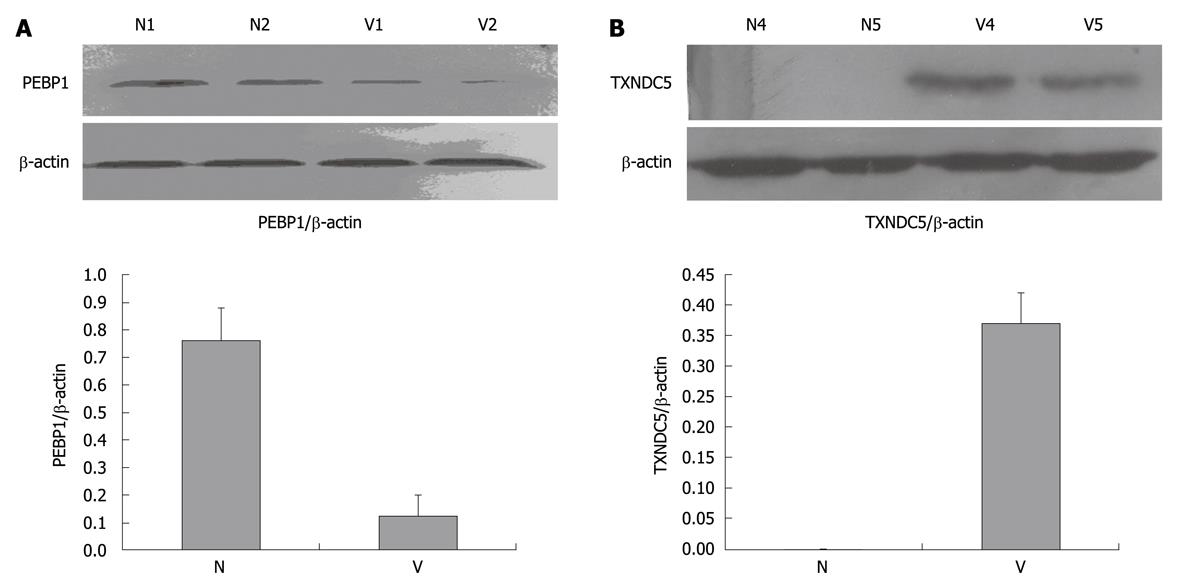
Figure 2 Western blotting analysis of phosphatidylethanolamine-binding protein 1 and thioredoxin domain-containing protein 5.
A: Marked downregulation of phosphatidylethanolamine-binding protein 1 (PEBP1) in varioliform gastritis (V) tissue. Protein extracts (50 μg) were separated on a 12% sodium dodecyl sulfate-polyacrylamide gel. Proteins were transferred to a poly-vinylidene difluoride membrane. After blocking, the membranes were incubated with rabbit monoclonal antibody of PEBP1 (dilution of 1:2000) and subsequently incubated with HRP-anti-rabbit IgG. The specific proteins were visualized with chemiluminescent reagent. As a control for equal protein loading, blots were restained with anti-actin antibody. Immunosignals were quantified by densitometry scanning. The relative quantification was calculated as the ratio of PEBP1 expression to actin expression as shown in the followed chart; B: Upregulation of thioredoxin domain-containing protein 5 (TXNDC5) in varioliform gastritis in comparison with that in normal (N) mucosa. The same experimental process was performed, except that the membranes were incubated with polyclonal goat anti-TXNDC5 antibody (dilution of 1:1000).
-
Citation: Zhang L, Hou YH, Wu K, Zhai JS, Lin N. Proteomic analysis reveals molecular biological details in varioliform gastritis without
Helicobacter pylori infection. World J Gastroenterol 2010; 16(29): 3664-3673 - URL: https://www.wjgnet.com/1007-9327/full/v16/i29/3664.htm
- DOI: https://dx.doi.org/10.3748/wjg.v16.i29.3664


