Copyright
©2010 Baishideng.
World J Gastroenterol. Aug 7, 2010; 16(29): 3642-3650
Published online Aug 7, 2010. doi: 10.3748/wjg.v16.i29.3642
Published online Aug 7, 2010. doi: 10.3748/wjg.v16.i29.3642
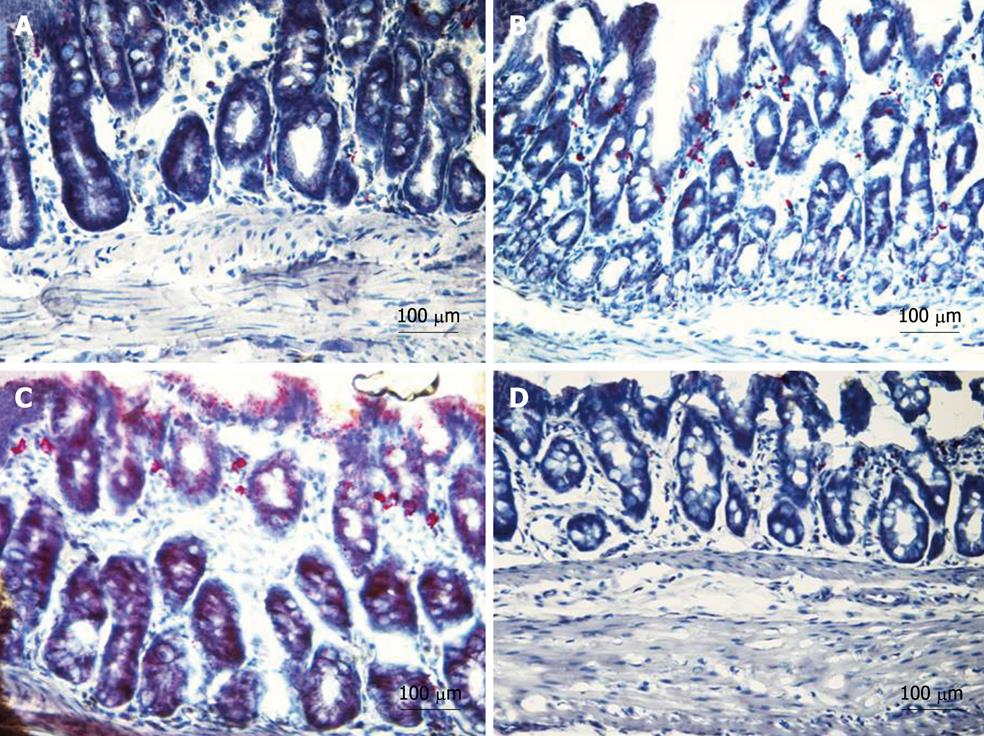
Figure 6 Naphthol AS-D chloroacetate esterase positivity (red) in cross section of distal colon.
A: Tissue sections obtained from non-inflamed rats showed occasional red staining indicating a low presence of neutrophils within the bowel wall under physiological conditions; B: From a rat with colitis (5% dextran sodium sulphate in drinking water, panel B). Compared to non-inflamed rats, tissue from rats with colitis showed a massive neutrophil infiltration extending throughout the mucosa (note the scattered degranulation within the crypts and submucosa); C, D: Treatment with bis(1-hydroxy-2,2,6,6-tetramethyl-4-piperidinyl)decandioate (IAC) 30 mg/kg hydrophilic form po (C) was unable to suppress neutrophil infiltration especially in the mucosa. Treatment with lipophilic IAC both po (not shown) and ip (D) almost completely suppressed neutrophil infiltration within the colonic wall.
- Citation: Vasina V, Broccoli M, Ursino MG, Canistro D, Valgimigli L, Soleti A, Paolini M, Ponti FD. Non-peptidyl low molecular weight radical scavenger IAC attenuates DSS-induced colitis in rats. World J Gastroenterol 2010; 16(29): 3642-3650
- URL: https://www.wjgnet.com/1007-9327/full/v16/i29/3642.htm
- DOI: https://dx.doi.org/10.3748/wjg.v16.i29.3642


