Copyright
©2010 Baishideng.
World J Gastroenterol. Aug 7, 2010; 16(29): 3642-3650
Published online Aug 7, 2010. doi: 10.3748/wjg.v16.i29.3642
Published online Aug 7, 2010. doi: 10.3748/wjg.v16.i29.3642
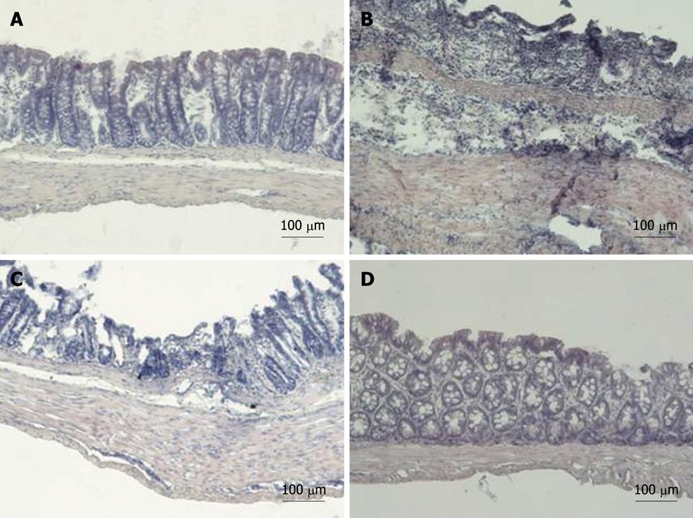
Figure 5 Representative examples of cross sections of distal colon.
A: From a non-inflamed rat [drinking water + bis(1-hydroxy-2,2,6,6-tetramethyl-4-piperidinyl)decandioate (IAC) vehicle orally]; B: From an inflamed rat [dextran sodium sulphate (DSS) 5% in drinking water + IAC vehicle orally]. Note the dramatic loss of mucosal architecture with crypt dropout and the granulocyte infiltrate extending throughout the mucosa and submucosa; C, D: Cross sections of distal colon from an inflamed rat treated with hydrophilic (C) and lipophilic (D) IAC 30 mg/kg orally (C) and intraperitoneally (D). Lipophilic (po, not shown here, and ip) IAC 30 mg/kg decreased the microscopic damage produced by DSS, facilitating mucosal healing, reducing inflammatory cells infiltration and muscle thickening (panel D). Hydrophilic IAC po failed to protect the colon from the damage induced by DSS (panel C).
- Citation: Vasina V, Broccoli M, Ursino MG, Canistro D, Valgimigli L, Soleti A, Paolini M, Ponti FD. Non-peptidyl low molecular weight radical scavenger IAC attenuates DSS-induced colitis in rats. World J Gastroenterol 2010; 16(29): 3642-3650
- URL: https://www.wjgnet.com/1007-9327/full/v16/i29/3642.htm
- DOI: https://dx.doi.org/10.3748/wjg.v16.i29.3642


