Copyright
©2010 Baishideng.
World J Gastroenterol. Jul 14, 2010; 16(26): 3292-3298
Published online Jul 14, 2010. doi: 10.3748/wjg.v16.i26.3292
Published online Jul 14, 2010. doi: 10.3748/wjg.v16.i26.3292
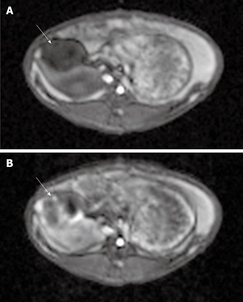
Figure 4 T1-weighted gradient-echo magnetic resonance [taken before (A) and after (B) TRIP imaging occurred].
Each image shows a VX2 tumor (solid arrows) located in the pancreas. Note the areas of increased perfusion to the viable tumor periphery in image B.
- Citation: Lewandowski RJ, Eifler AC, Bentrem DJ, Chung JC, Wang D, Woloschak GE, Yang GY, Ryu R, Salem R, Larson AC, Omary RA. Functional magnetic resonance imaging in an animal model of pancreatic cancer. World J Gastroenterol 2010; 16(26): 3292-3298
- URL: https://www.wjgnet.com/1007-9327/full/v16/i26/3292.htm
- DOI: https://dx.doi.org/10.3748/wjg.v16.i26.3292


