Copyright
©2010 Baishideng.
World J Gastroenterol. Jun 21, 2010; 16(23): 2954-2958
Published online Jun 21, 2010. doi: 10.3748/wjg.v16.i23.2954
Published online Jun 21, 2010. doi: 10.3748/wjg.v16.i23.2954
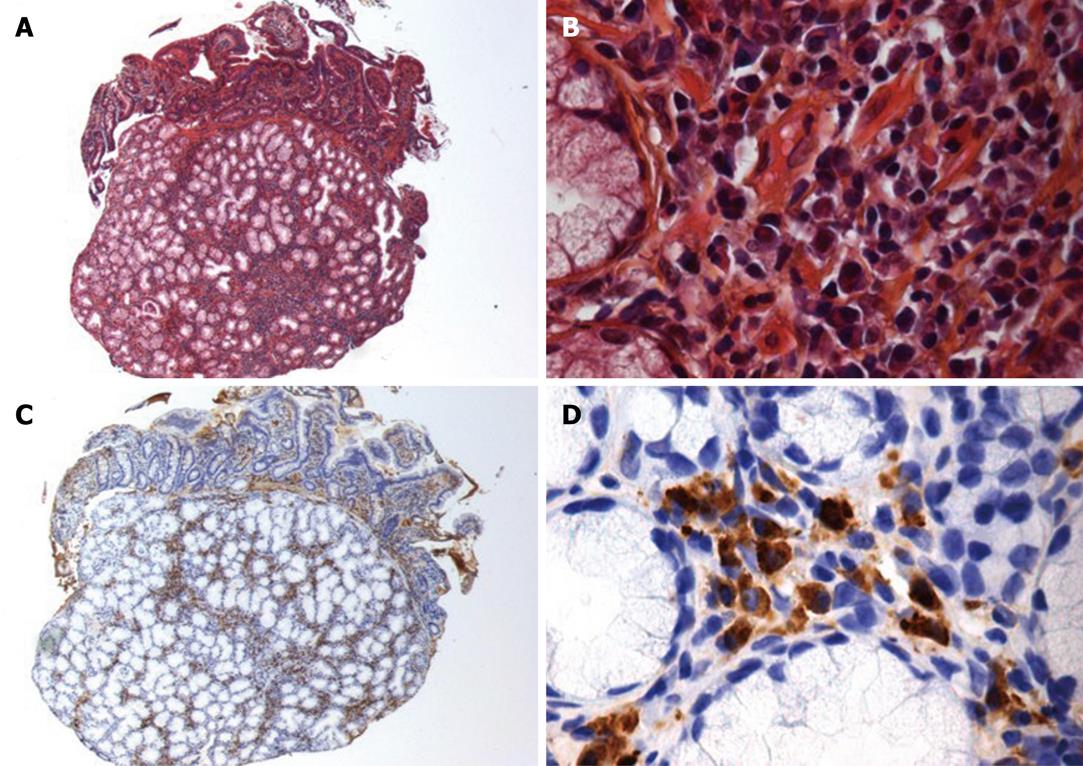
Figure 3 Histological examination of periampular biopsies (notice that both duodenal mucosa and pancreatic parenchyma are visible).
A, B: Hematoxylin-eosin staining (A: × 125, B: × 500): atrophic acini surrounded by a fibrous stroma, associated with periductal lymphoplasmocytic infiltration; C, D: Anti-IgG4 immunostaining (C: × 125, D: × 500): the plasma cell infiltration expresses IgG4.
- Citation: Neuzillet C, Lepère C, Hajjam ME, Palazzo L, Fabre M, Turki H, Hammel P, Rougier P, Mitry E. Autoimmune pancreatitis with atypical imaging findings that mimicked an endocrine tumor. World J Gastroenterol 2010; 16(23): 2954-2958
- URL: https://www.wjgnet.com/1007-9327/full/v16/i23/2954.htm
- DOI: https://dx.doi.org/10.3748/wjg.v16.i23.2954


