Copyright
©2010 Baishideng.
World J Gastroenterol. Jun 21, 2010; 16(23): 2954-2958
Published online Jun 21, 2010. doi: 10.3748/wjg.v16.i23.2954
Published online Jun 21, 2010. doi: 10.3748/wjg.v16.i23.2954
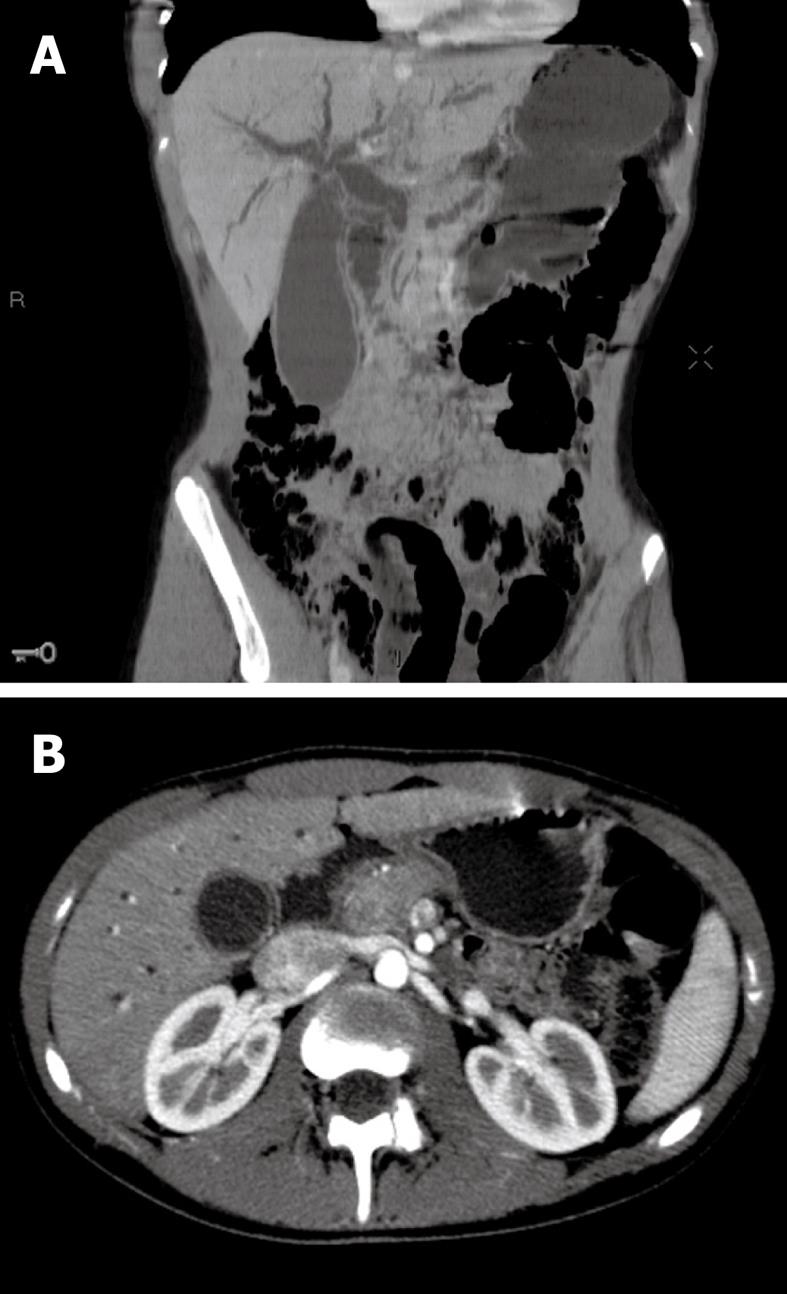
Figure 1 Initial multi-detector computed tomography scan imaging.
A: CT scan before contrast injection, coronal reconstruction: enlargement of the main pancreatic duct and the biliary ducts, with enlarged gallbladder; B: CT scan after contrast injection, arterial phase, axial slice: lesion of the head of the pancreas, moderately enhanced after contrast injection.
- Citation: Neuzillet C, Lepère C, Hajjam ME, Palazzo L, Fabre M, Turki H, Hammel P, Rougier P, Mitry E. Autoimmune pancreatitis with atypical imaging findings that mimicked an endocrine tumor. World J Gastroenterol 2010; 16(23): 2954-2958
- URL: https://www.wjgnet.com/1007-9327/full/v16/i23/2954.htm
- DOI: https://dx.doi.org/10.3748/wjg.v16.i23.2954


