Copyright
©2010 Baishideng.
World J Gastroenterol. Jun 21, 2010; 16(23): 2835-2840
Published online Jun 21, 2010. doi: 10.3748/wjg.v16.i23.2835
Published online Jun 21, 2010. doi: 10.3748/wjg.v16.i23.2835
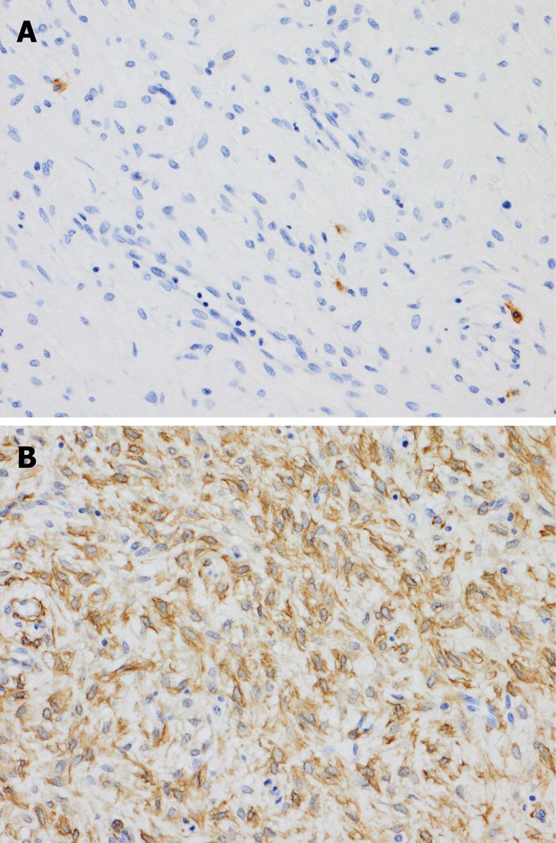
Figure 3 The results of immunohistochemical staining.
A: The tumor cells are negative for KIT. The scattered KIT-positive cells are mast cells (× 200); B: The tumor cells are diffusely positive for smooth muscle actin (× 200).
- Citation: Takahashi Y, Suzuki M, Fukusato T. Plexiform angiomyxoid myofibroblastic tumor of the stomach. World J Gastroenterol 2010; 16(23): 2835-2840
- URL: https://www.wjgnet.com/1007-9327/full/v16/i23/2835.htm
- DOI: https://dx.doi.org/10.3748/wjg.v16.i23.2835


