Copyright
©2010 Baishideng.
World J Gastroenterol. Jun 21, 2010; 16(23): 2835-2840
Published online Jun 21, 2010. doi: 10.3748/wjg.v16.i23.2835
Published online Jun 21, 2010. doi: 10.3748/wjg.v16.i23.2835
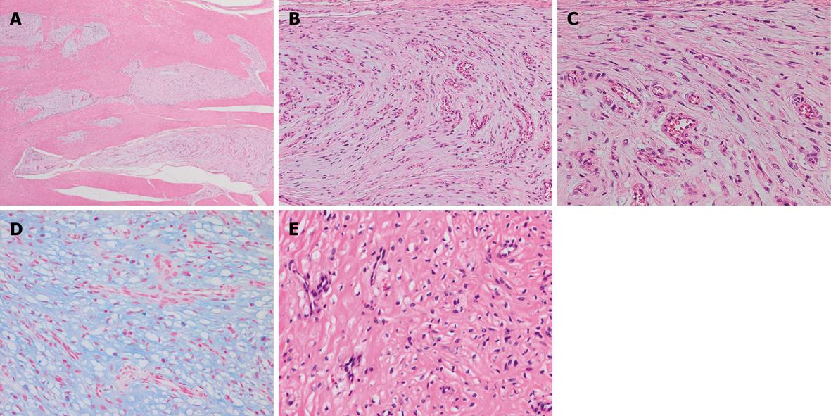
Figure 2 Histological appearance of PAMT.
A: The tumor shows a multinodular plexiform growth pattern (HE stain, × 20); B: Spindle-shaped bland tumor cells are separated by an abundant intercellular myxoid matrix. The stroma is rich in small vessels (HE stain, × 100); C: Tumor cells possess oval nuclei and a slightly eosinophilic cytoplasm. The nucleoli are inconspicuous and the cell borders are indistinct (HE stain, × 200); D: The stroma is positive for Alcian blue stain (× 200); E: Stromal collagenization is occasionally observed (HE stain, × 200).
- Citation: Takahashi Y, Suzuki M, Fukusato T. Plexiform angiomyxoid myofibroblastic tumor of the stomach. World J Gastroenterol 2010; 16(23): 2835-2840
- URL: https://www.wjgnet.com/1007-9327/full/v16/i23/2835.htm
- DOI: https://dx.doi.org/10.3748/wjg.v16.i23.2835


