Copyright
©2010 Baishideng.
World J Gastroenterol. Jun 14, 2010; 16(22): 2824-2827
Published online Jun 14, 2010. doi: 10.3748/wjg.v16.i22.2824
Published online Jun 14, 2010. doi: 10.3748/wjg.v16.i22.2824
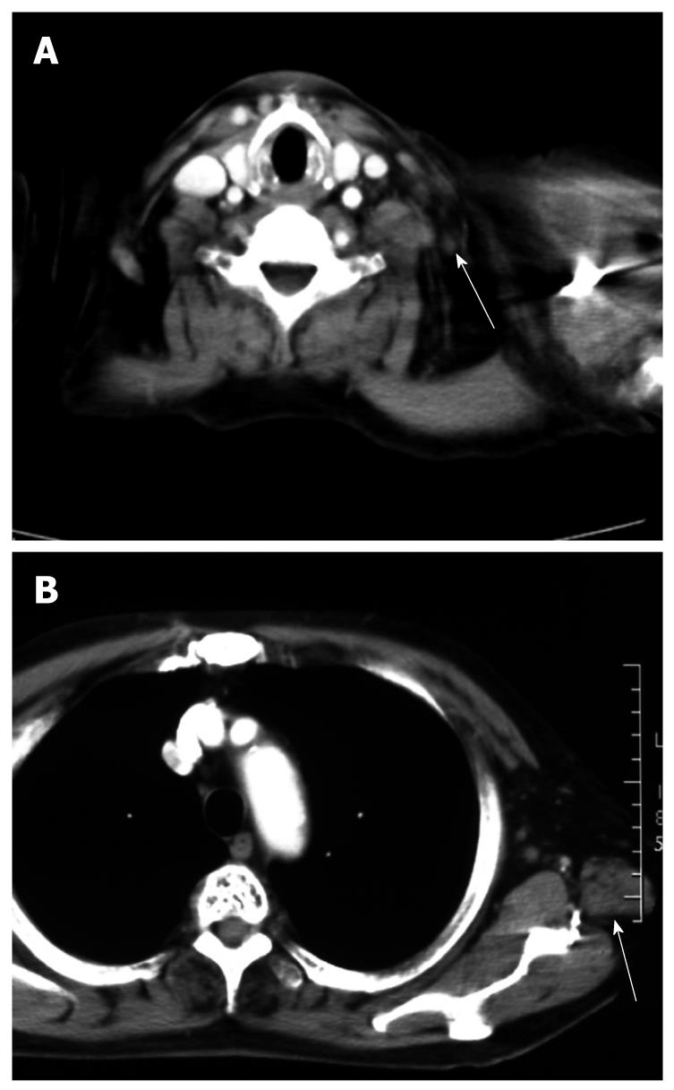
Figure 2 CT shows a Virchow’s (A) and an axillary lymph node (B) which were reduced remarkably in size after treatment.
- Citation: Hijioka S, Chin K, Seto Y, Yamamoto N, Hatake K. Eight-year survival after advanced gastric cancer treated with S-1 followed by surgery. World J Gastroenterol 2010; 16(22): 2824-2827
- URL: https://www.wjgnet.com/1007-9327/full/v16/i22/2824.htm
- DOI: https://dx.doi.org/10.3748/wjg.v16.i22.2824


