Copyright
©2010 Baishideng.
World J Gastroenterol. Jun 14, 2010; 16(22): 2743-2753
Published online Jun 14, 2010. doi: 10.3748/wjg.v16.i22.2743
Published online Jun 14, 2010. doi: 10.3748/wjg.v16.i22.2743
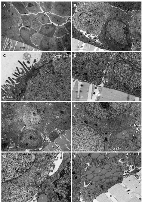
Figure 3 Undifferentiated heterogenous population of HT-29 cells.
A-D: HT-29 cells grown on membrane filters (mem) and examined day 1 post-confluency. A: Stratified cuboidal cells arranged in clumps. Large intercellular spaces (black arrows) are evident with microvilli projecting into them; B: High power micrograph showing large intercellular spaces (approximate 2 μm) (white arrows) with microvillar projections and formation of small vacuoles (v); C: Microvillous (m) apical plasma membrane is present and desmosomes (*) are evident along contact sites of the lateral plasma membrane of adjoining cells and presence of intercellular spaces (white arrow); D: No invasive projections are evident along the basolateral plasma membrane. Desmosomes (*) are present at contact sites of the plasma membrane, which otherwise shows intercellular spaces (white arrow); E-H: HT-29 cells grown on membrane filters (mem) and examined day 12 post-confluency. E: Stratified cuboidal cells with microvillar projections entering the large intercellular space (black arrow). Intranuclear rods are evident (inr); F: High power micrograph of junction between two cells. Golgi bodies (g) as well as mucin filled vacuoles (v) are distributed throughout the cell cytoplasm and intercellular spaces are evident with microvilli projecting into their lumen; G: High power micrograph of a contact site between two opposing plasma membranes showing a desmosome (*). Large intercellular spaces (black arrows) are evident above and below the desmosomes contact site. Mitochondria (mit) and mucin filled vacuoles (v) are also evident; H: Basolateral attachment site showing minor invasive projections (inv) into the filter pore. Intercellular spaces (black arrow) and vacuoles (v) are evident.
-
Citation: Biazik JM, Jahn KA, Su Y, Wu YN, Braet F. Unlocking the ultrastructure of colorectal cancer cells
in vitro using selective staining. World J Gastroenterol 2010; 16(22): 2743-2753 - URL: https://www.wjgnet.com/1007-9327/full/v16/i22/2743.htm
- DOI: https://dx.doi.org/10.3748/wjg.v16.i22.2743


