Copyright
©2010 Baishideng.
World J Gastroenterol. Jun 14, 2010; 16(22): 2743-2753
Published online Jun 14, 2010. doi: 10.3748/wjg.v16.i22.2743
Published online Jun 14, 2010. doi: 10.3748/wjg.v16.i22.2743
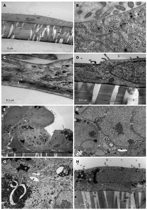
Figure 1 Spontaneous differentiation of Caco-2 into sub-population 1 of stratified cuboidal cells.
A-D: Caco-2 cells grown on membrane filters examined on day 1 one post-confluency. A: Simple squamous cells with irregular sparse microvilli; B: Junction between two cells (black arrow) and establishment of a tight junction (white arrow) and the presence of microvilli (m) and golgi bodies (g); C: Desmosome (*) formation along the lateral plasma membrane between two cells (white arrow); D: Basolateral plasma membrane depicting attachment to membrane filter with evidence of small cell protrusions (inv) into the membrane pore (p). Mitochondria (mit); E-H: Caco-2 cells grown on membrane filters examined 12 d post-confluency. E: Cells appear stratified and cuboidal with large intercellular spaces (black arrows); large glycogen stores are evident (gl); F: Intercellular spaces (black arrows) and desmosomes (*) are evident between cells with glycogen deposits (gl) and indentations of the nucleus form large intranuclear rods (inr); G: High power image of intercellular spaces (black arrows) and desmosomes (*) present along the lateral plasma membrane with large glycogen deposits (gl) found in the cytoplasm; H: Basolateral plasma membrane showing attachment of the cell to the membrane filter. No invasive processes are evident projecting into the filter pores (p). Glycogen (gl) and some lipid (lp) droplets are present in the cell cytoplasm.
-
Citation: Biazik JM, Jahn KA, Su Y, Wu YN, Braet F. Unlocking the ultrastructure of colorectal cancer cells
in vitro using selective staining. World J Gastroenterol 2010; 16(22): 2743-2753 - URL: https://www.wjgnet.com/1007-9327/full/v16/i22/2743.htm
- DOI: https://dx.doi.org/10.3748/wjg.v16.i22.2743


