Copyright
©2010 Baishideng.
World J Gastroenterol. Jun 14, 2010; 16(22): 2735-2742
Published online Jun 14, 2010. doi: 10.3748/wjg.v16.i22.2735
Published online Jun 14, 2010. doi: 10.3748/wjg.v16.i22.2735
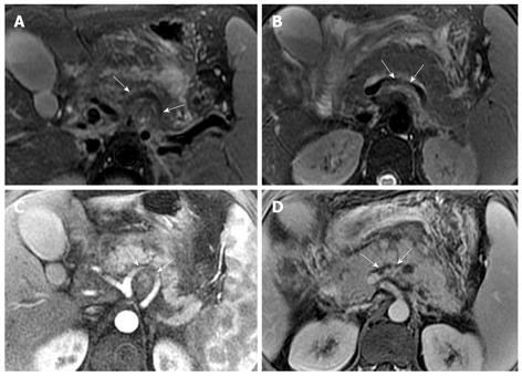
Figure 11 30-year-old man with acute pancreatitis and splenomegaly.
Axial T2-weighted images (A and B) show the involved segments (arrows) of splenic artery (A) and splenic vein (B). Enhanced arterial phase (C) and venous phase (D) images reveal clearly the involved segments (arrows) of the splenic artery (C) and vein (D).
- Citation: Xiao B, Zhang XM, Tang W, Zeng NL, Zhai ZH. Magnetic resonance imaging for local complications of acute pancreatitis: A pictorial review. World J Gastroenterol 2010; 16(22): 2735-2742
- URL: https://www.wjgnet.com/1007-9327/full/v16/i22/2735.htm
- DOI: https://dx.doi.org/10.3748/wjg.v16.i22.2735


