Copyright
©2010 Baishideng.
World J Gastroenterol. Jun 7, 2010; 16(21): 2669-2676
Published online Jun 7, 2010. doi: 10.3748/wjg.v16.i21.2669
Published online Jun 7, 2010. doi: 10.3748/wjg.v16.i21.2669
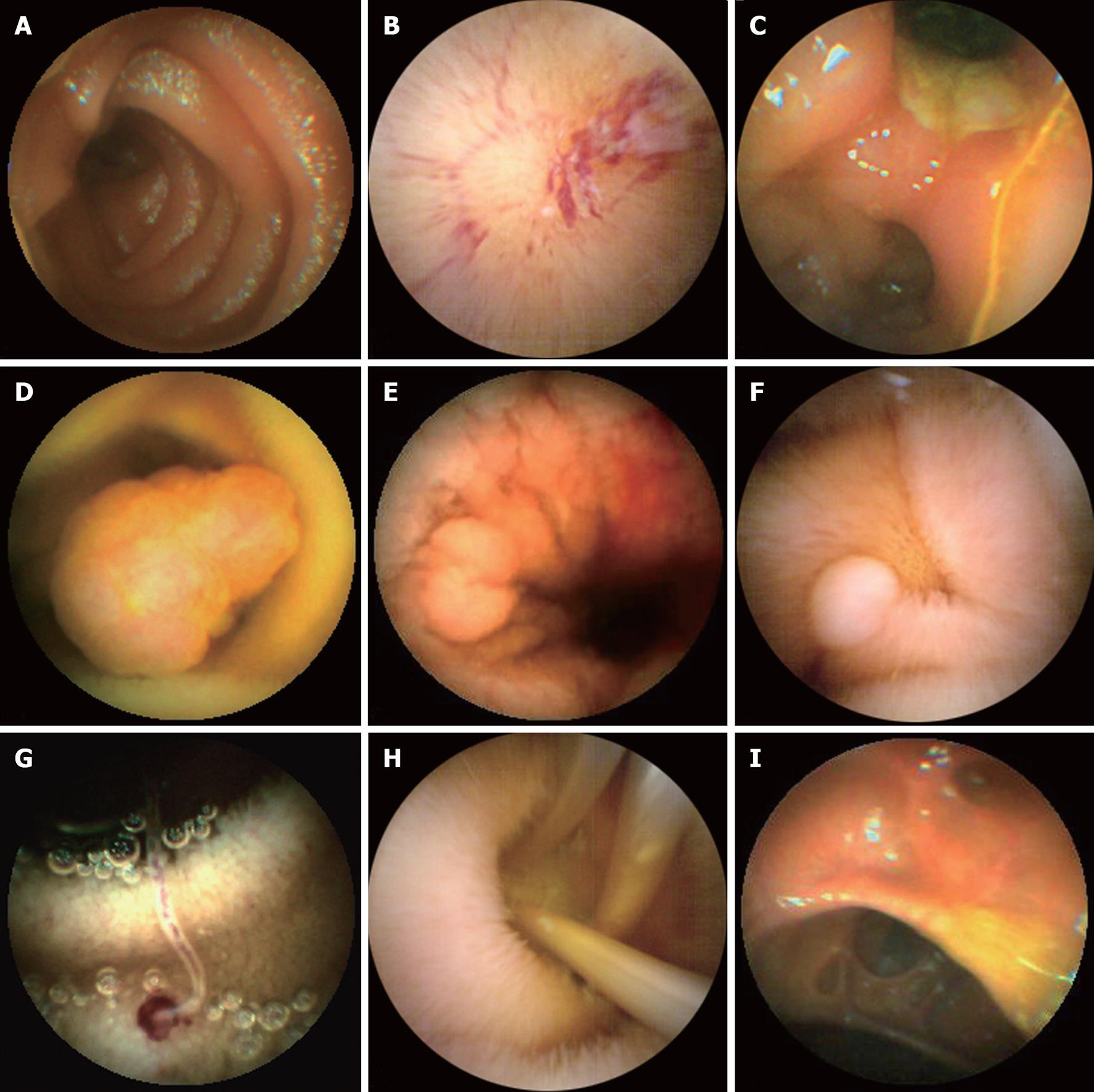
Figure 2 Small bowel images captured by the OMOM capsule endoscope.
A: Normal small bowel mucosa; B: Angiectasias; C: Ulcer; D: Tumor; E: Crohn’s disease; F: Polyp; G: Hookworm; H: Lumbricoides; I: Multiple diverticulum.
- Citation: Liao Z, Gao R, Li F, Xu C, Zhou Y, Wang JS, Li ZS. Fields of applications, diagnostic yields and findings of OMOM capsule endoscopy in 2400 Chinese patients. World J Gastroenterol 2010; 16(21): 2669-2676
- URL: https://www.wjgnet.com/1007-9327/full/v16/i21/2669.htm
- DOI: https://dx.doi.org/10.3748/wjg.v16.i21.2669


