Copyright
©2010 Baishideng.
World J Gastroenterol. May 28, 2010; 16(20): 2566-2570
Published online May 28, 2010. doi: 10.3748/wjg.v16.i20.2566
Published online May 28, 2010. doi: 10.3748/wjg.v16.i20.2566
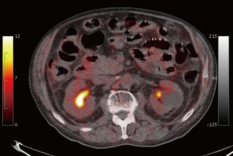
Figure 5 18FDG-positron emission tomography (PET)/CT images did not show focal or diffuse increase of the tracer uptake of small bowel walls.
- Citation: Mainenti PP, Segreto S, Mancini M, Rispo A, Cozzolino I, Masone S, Rinaldi CR, Nardone G, Salvatore M. Intestinal amyloidosis: Two cases with different patterns of clinical and imaging presentation. World J Gastroenterol 2010; 16(20): 2566-2570
- URL: https://www.wjgnet.com/1007-9327/full/v16/i20/2566.htm
- DOI: https://dx.doi.org/10.3748/wjg.v16.i20.2566


