Copyright
©2010 Baishideng.
World J Gastroenterol. May 28, 2010; 16(20): 2566-2570
Published online May 28, 2010. doi: 10.3748/wjg.v16.i20.2566
Published online May 28, 2010. doi: 10.3748/wjg.v16.i20.2566
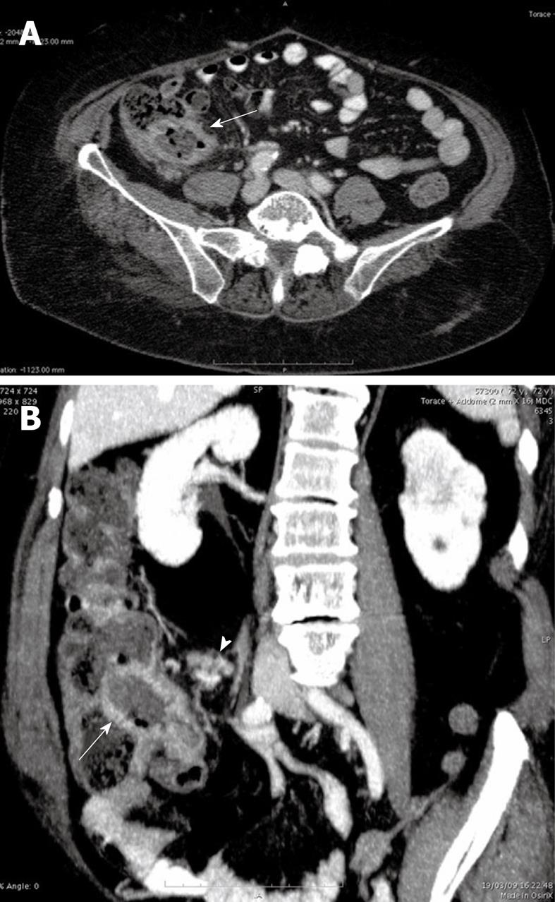
Figure 1 The axial computed tomography (CT) image (A) and the multiplanar reformatted (MPR) coronal oblique image (B) showed thickened walls of the terminal ileum with mild pseudoaneurysmal dilatation of the lumen (arrows) and multiple ileo-cecal lymph nodes (arrow head).
- Citation: Mainenti PP, Segreto S, Mancini M, Rispo A, Cozzolino I, Masone S, Rinaldi CR, Nardone G, Salvatore M. Intestinal amyloidosis: Two cases with different patterns of clinical and imaging presentation. World J Gastroenterol 2010; 16(20): 2566-2570
- URL: https://www.wjgnet.com/1007-9327/full/v16/i20/2566.htm
- DOI: https://dx.doi.org/10.3748/wjg.v16.i20.2566


