Copyright
©2010 Baishideng.
World J Gastroenterol. May 28, 2010; 16(20): 2504-2519
Published online May 28, 2010. doi: 10.3748/wjg.v16.i20.2504
Published online May 28, 2010. doi: 10.3748/wjg.v16.i20.2504
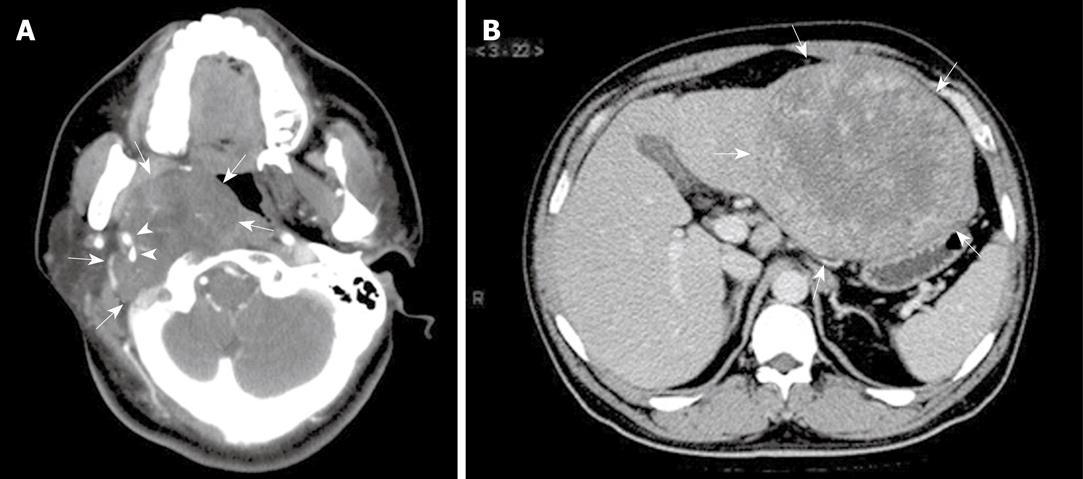
Figure 1 Radiological features of extranodal follicular dendritic cell (FDC) sarcoma shown by computed tomographic scan.
A: Case 8, a tumor (arrows) at the right parapharyngeal space showing soft tissue-like density and an expansive growth pattern, with the internal and external carotid arteries (arrowheads) engulfed; B: Case 6, a well-circumscribed mass (arrows) at the left lobe of liver, showing irregular enhancement at its periphery.
- Citation: Li L, Shi YH, Guo ZJ, Qiu T, Guo L, Yang HY, Zhang X, Zhao XM, Su Q. Clinicopathological features and prognosis assessment of extranodal follicular dendritic cell sarcoma. World J Gastroenterol 2010; 16(20): 2504-2519
- URL: https://www.wjgnet.com/1007-9327/full/v16/i20/2504.htm
- DOI: https://dx.doi.org/10.3748/wjg.v16.i20.2504


