Copyright
©2010 Baishideng.
World J Gastroenterol. May 21, 2010; 16(19): 2341-2347
Published online May 21, 2010. doi: 10.3748/wjg.v16.i19.2341
Published online May 21, 2010. doi: 10.3748/wjg.v16.i19.2341
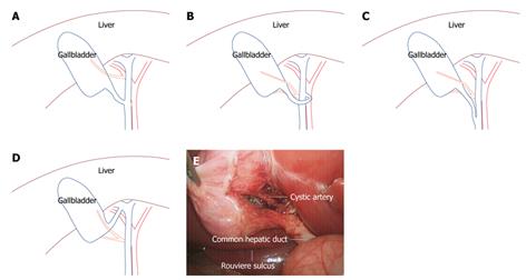
Figure 1 Anatomy of laparoscopic cholecystectomy (LC) (blue curves stand for bile duct and gallbladder and red curves for arteries).
A: Illustration of a variant cystic artery originating from the right hepatic artery; B: Cystic duct connected to the left wall of hepatic duct via an anterior approach; C: Cystic duct parallel with common hepatic duct; D: Cystic duct connected to the right hepatic duct; E: Calot’s triangle and Rouviere sulcus in LC.
- Citation: Li LJ, Zheng XM, Jiang DZ, Zhang W, Shen HL, Shan CX, Liu S, Qiu M. Progress in laparoscopic anatomy research: A review of the Chinese literature. World J Gastroenterol 2010; 16(19): 2341-2347
- URL: https://www.wjgnet.com/1007-9327/full/v16/i19/2341.htm
- DOI: https://dx.doi.org/10.3748/wjg.v16.i19.2341


