Copyright
©2010 Baishideng.
World J Gastroenterol. May 14, 2010; 16(18): 2305-2310
Published online May 14, 2010. doi: 10.3748/wjg.v16.i18.2305
Published online May 14, 2010. doi: 10.3748/wjg.v16.i18.2305
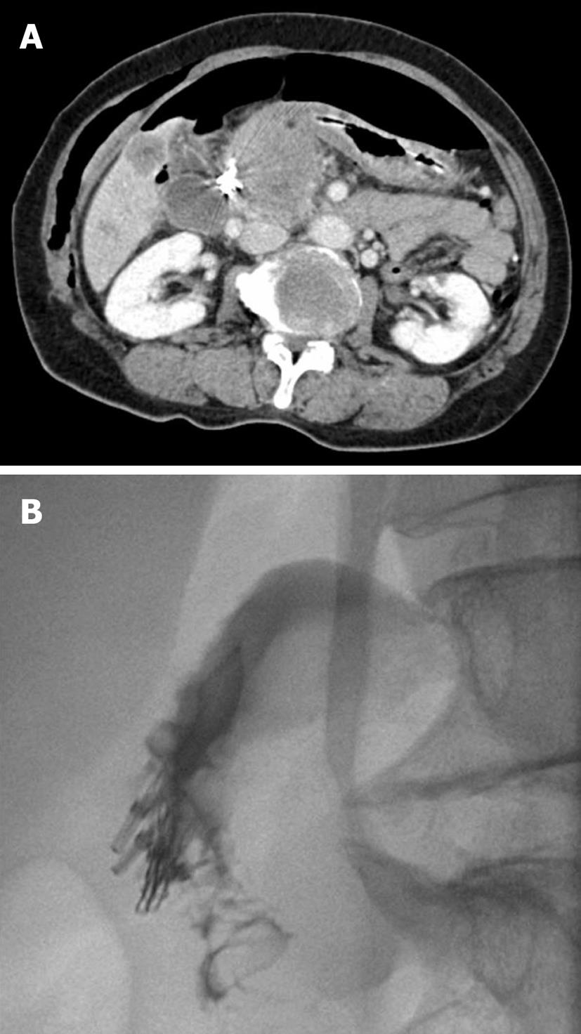
Figure 4 Initial abdominal CT following perforation shows pneumoperitoneum and subcutaneous emphysema (A), and follow up UGIs performed 8 d later reveal no contrast leaks (B).
- Citation: Lee TH, Bang BW, Jeong JI, Kim HG, Jeong S, Park SM, Lee DH, Park SH, Kim SJ. Primary endoscopic approximation suture under cap-assisted endoscopy of an ERCP-induced duodenal perforation. World J Gastroenterol 2010; 16(18): 2305-2310
- URL: https://www.wjgnet.com/1007-9327/full/v16/i18/2305.htm
- DOI: https://dx.doi.org/10.3748/wjg.v16.i18.2305


