Copyright
©2010 Baishideng.
World J Gastroenterol. May 14, 2010; 16(18): 2291-2297
Published online May 14, 2010. doi: 10.3748/wjg.v16.i18.2291
Published online May 14, 2010. doi: 10.3748/wjg.v16.i18.2291
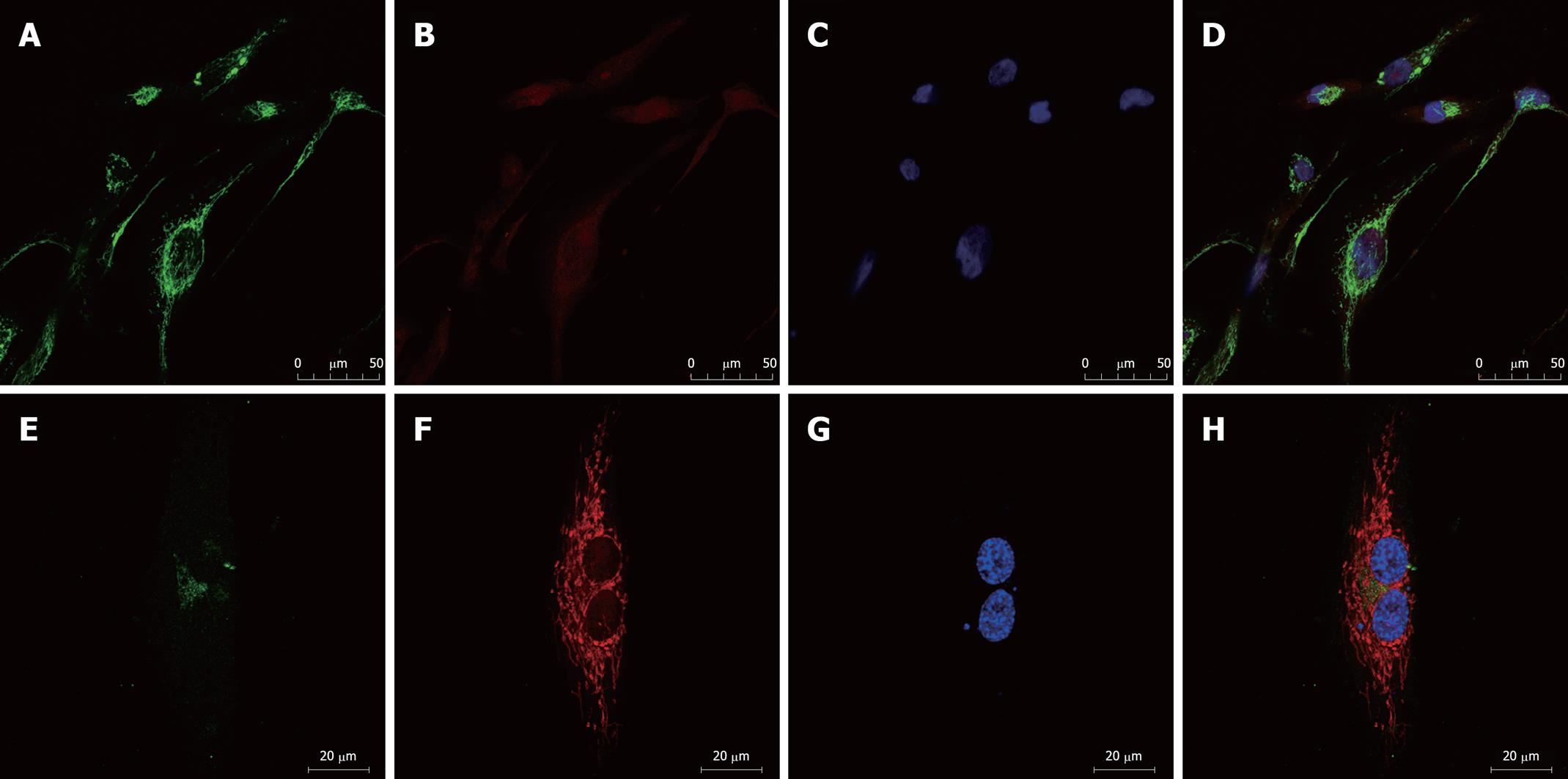
Figure 3 Laser scanning confocal microscopy for MDR1/P-gp in resistance SK-Hep-1/CDDP (A-D) and parent SK-Hep-1 (E-H) cell lines.
SK-Hep-1/CDDP (A) and SK-Hep-1 (E) cells were simultaneously stained for the anti-P-gp antibody-FITC (green), (B) and (F) were stained with mitochondria (red), (C) and (G) immunofluorescence images for the nuclei (blue), (D) and (H) the expression of the merged image. MDR1: Multidrug resistant protein 1; P-gp: Phospho-glycoprotein.
-
Citation: Zhou Y, Ling XL, Li SW, Li XQ, Yan B. Establishment of a human hepatoma multidrug resistant cell line
in vitro . World J Gastroenterol 2010; 16(18): 2291-2297 - URL: https://www.wjgnet.com/1007-9327/full/v16/i18/2291.htm
- DOI: https://dx.doi.org/10.3748/wjg.v16.i18.2291


