Copyright
©2010 Baishideng.
World J Gastroenterol. May 7, 2010; 16(17): 2100-2108
Published online May 7, 2010. doi: 10.3748/wjg.v16.i17.2100
Published online May 7, 2010. doi: 10.3748/wjg.v16.i17.2100
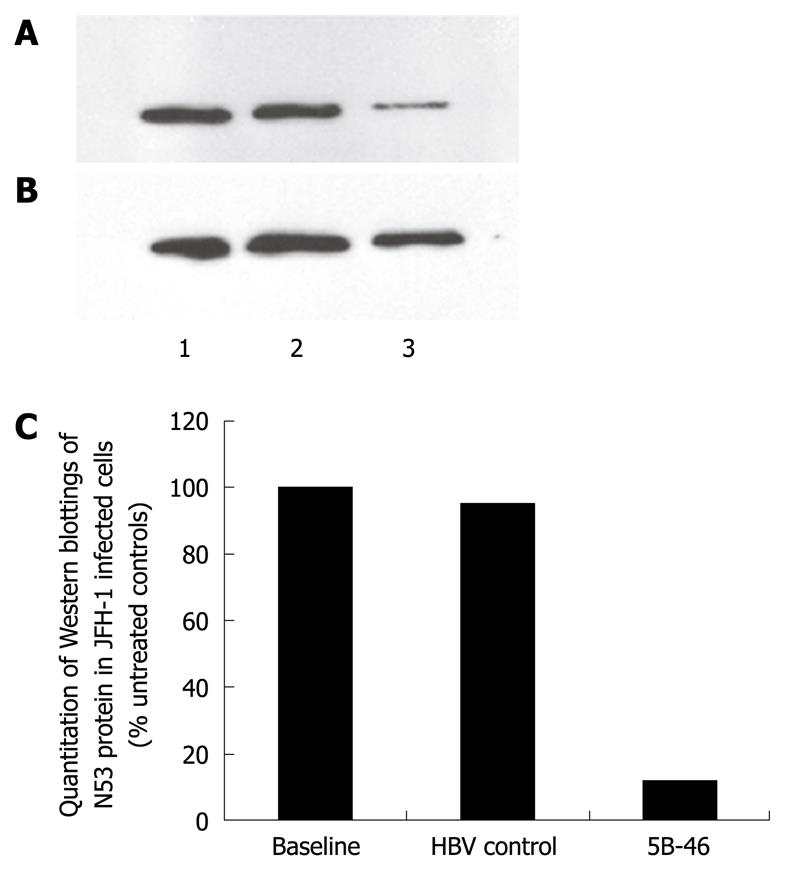
Figure 5 Western blotting analysis of NS3 protease gene expression in JFH-1 infected Huh7.
5 cells after transfection of 5B-74 mimic and controls. A: HCV NS3; B: Tubulin, 72 h after transfection. Lane 1: Levels prior to addition of mimic; Lane 2: HBV (unrelated) control; Lane 3: 5B-46 mimic; C: Quantification of Western blottings of NS3 protein in JFH-1-infected cells at baseline, after 72 h exposure to 5B-46, and to HBV unrelated control. Bound NS3 antibodies were detected with SuperSignal chemiluminescent substrate and quantitated by Gel Imaging software GeneTools.
- Citation: Smolic R, Smolic M, Andorfer JH, Wu CH, Smith RM, Wu GY. Inhibition of hepatitis C virus replication by single-stranded RNA structural mimics. World J Gastroenterol 2010; 16(17): 2100-2108
- URL: https://www.wjgnet.com/1007-9327/full/v16/i17/2100.htm
- DOI: https://dx.doi.org/10.3748/wjg.v16.i17.2100


