Copyright
©2010 Baishideng.
World J Gastroenterol. Apr 28, 2010; 16(16): 2038-2045
Published online Apr 28, 2010. doi: 10.3748/wjg.v16.i16.2038
Published online Apr 28, 2010. doi: 10.3748/wjg.v16.i16.2038
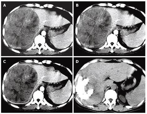
Figure 2 Transverse CT scans showing a massive pattern of HCC in the right lobe of a 46-year-old man with a tumor volume of 1936.
70 cm3, a TTLVR of 67.52%, and a survival time of 6 mo. A and B: Arterial phase contrast- enhanced CT scan showing a high density tumor in the right lobe of liver before the first TACE procedure, while one-dimensional measurement showing the largest tumor diameter (17.7 cm) and two-dimensional measurement showing the tumor product of the greatest axial dimension (237.18 cm2); C: Contrast-enhanced CT scan showing hepatic lesion volumetric measurements before TACE; D: Nonenhanced CT follow-up scan showing lipiodol retention of grade III and a reduced tumor size 60 d after the first TACE procedure.
- Citation: Zhang JW, Feng XY, Liu HQ, Yao ZW, Yang YM, Liu B, Yu YQ. CT volume measurement for prognostic evaluation of unresectable hepatocellular carcinoma after TACE. World J Gastroenterol 2010; 16(16): 2038-2045
- URL: https://www.wjgnet.com/1007-9327/full/v16/i16/2038.htm
- DOI: https://dx.doi.org/10.3748/wjg.v16.i16.2038


