Copyright
©2010 Baishideng.
World J Gastroenterol. Apr 28, 2010; 16(16): 1970-1978
Published online Apr 28, 2010. doi: 10.3748/wjg.v16.i16.1970
Published online Apr 28, 2010. doi: 10.3748/wjg.v16.i16.1970
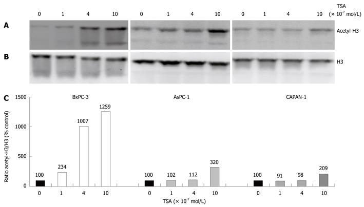
Figure 1 Enhancement of histone H3 acetylation by trichostatin-A (TSA).
The indicated pancreatic cancer (PC) cell lines were treated with various concentrations of TSA for 24 h. A: Histone H3 acetylation was analyzed by immunoblotting; B: Reprobing of the blot with an anti-H3 protein-specific antibody revealed no systematic differences of the histone H3 amount among the samples; C: Acetyl-histone H3 levels were further investigated using image analysis software and related to the histone H3 protein level. Therefore, acetyl-histone H3 and H3 protein signal intensities were determined, and the ratio acetyl-histone H3/H3 protein was calculated. A ratio of 100% corresponds to control cells cultured without TSA. The data shown are representative of three independent experiments.
- Citation: Emonds E, Fitzner B, Jaster R. Molecular determinants of the antitumor effects of trichostatin A in pancreatic cancer cells. World J Gastroenterol 2010; 16(16): 1970-1978
- URL: https://www.wjgnet.com/1007-9327/full/v16/i16/1970.htm
- DOI: https://dx.doi.org/10.3748/wjg.v16.i16.1970


