Copyright
©2010 Baishideng.
World J Gastroenterol. Apr 28, 2010; 16(16): 1953-1969
Published online Apr 28, 2010. doi: 10.3748/wjg.v16.i16.1953
Published online Apr 28, 2010. doi: 10.3748/wjg.v16.i16.1953
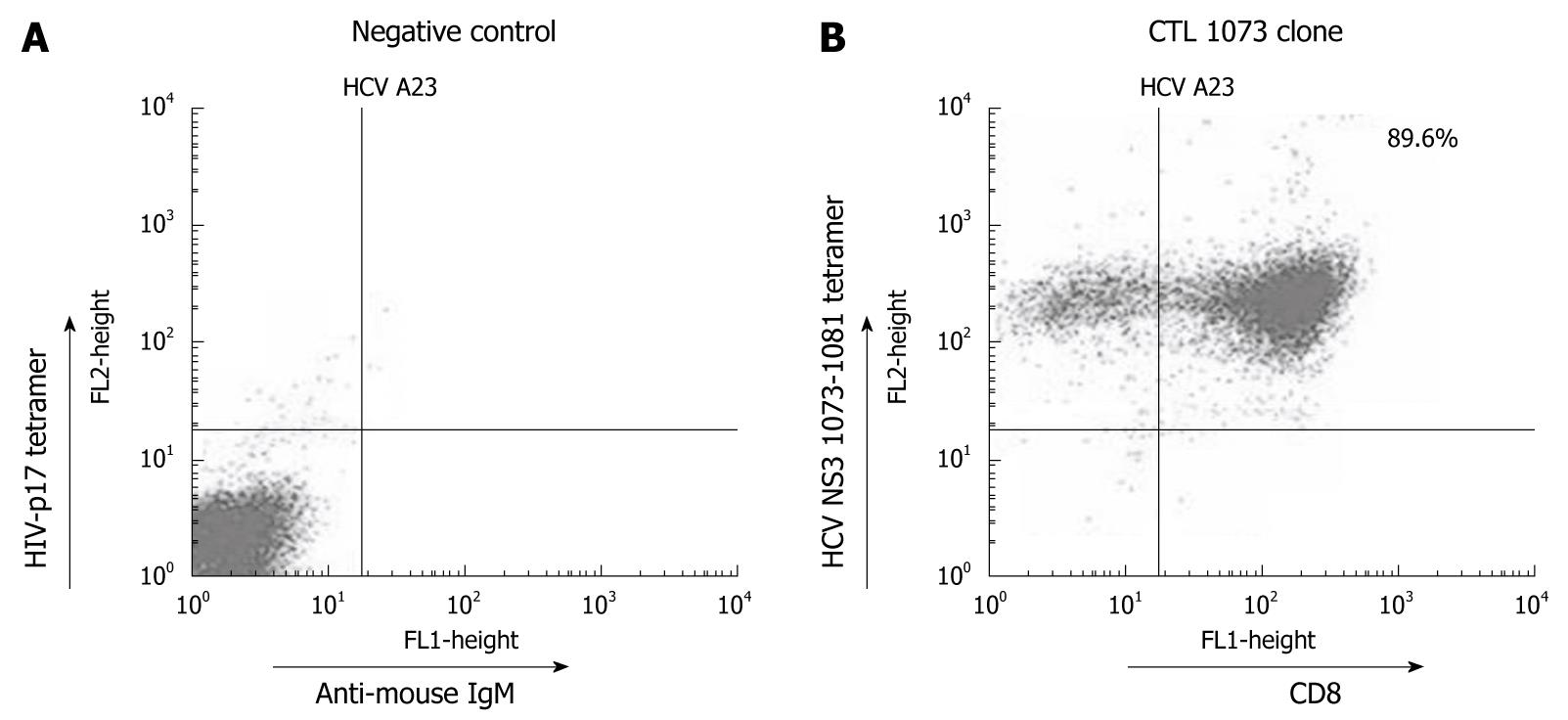
Figure 3 HCV NS3 1073-1081 tetramer staining using a CTL clone specific for wt1073.
A: As a negative control, CTL clone 1073 was stained with the HIV-p17 tetramer reagent and anti-mouse IgM FITC. As expected, no cell shift was observed; B: The same CTL clone was stained with the HCV NS3 1073-1081 tetramer and anti-CD8+-FITC. A positive HCV NS3 1073-1081 tetramer staining is shown with tetramer/CD8+ cells located in the upper right quadrant. The origin of cells seen in the upper left quadrant could not be identified and need further investigation. All staining experiments were repeated three times with similar results. Identical results were obtained with HCV NS3 1073-1081 tetramer staining using other CTL clones for wt1073 (data not shown).
- Citation: Wang S, Buchli R, Schiller J, Gao J, VanGundy RS, Hildebrand WH, Eckels DD. Natural epitope variants of the hepatitis C virus impair cytotoxic T lymphocyte activity. World J Gastroenterol 2010; 16(16): 1953-1969
- URL: https://www.wjgnet.com/1007-9327/full/v16/i16/1953.htm
- DOI: https://dx.doi.org/10.3748/wjg.v16.i16.1953


