Copyright
©2010 Baishideng.
World J Gastroenterol. Apr 14, 2010; 16(14): 1720-1726
Published online Apr 14, 2010. doi: 10.3748/wjg.v16.i14.1720
Published online Apr 14, 2010. doi: 10.3748/wjg.v16.i14.1720
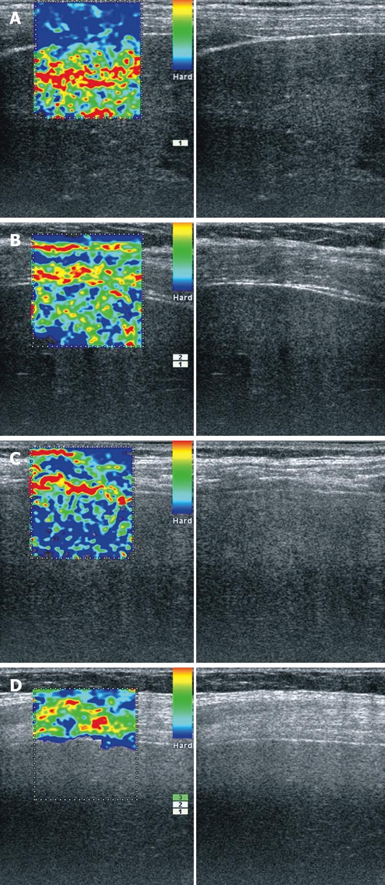
Figure 1 Real-time elastography images of right liver lobe.
A: 58-year-old patient with alcoholic liver steatosis - a very soft liver parenchyma (red/yellow/green) in contrast with hard intercostals muscles and diaphragm (blue); B: 56-year-old patient with chronic hepatitis C - parenchyma with mixed appearance (green/blue) indicative of elasticity; C: 57-year-old patient with alcoholic cirrhosis - very hard liver parenchyma (predominantly blue); D: 67-year-old obese woman with chronic hepatitis. The elastography software was not able to characterize elasticity inside liver.
- Citation: Gheonea DI, Săftoiu A, Ciurea T, Gorunescu F, Iordache S, Popescu GL, Belciug S, Gorunescu M, Săndulescu L. Real-time sono-elastography in the diagnosis of diffuse liver diseases. World J Gastroenterol 2010; 16(14): 1720-1726
- URL: https://www.wjgnet.com/1007-9327/full/v16/i14/1720.htm
- DOI: https://dx.doi.org/10.3748/wjg.v16.i14.1720


