Copyright
©2010 Baishideng.
World J Gastroenterol. Mar 28, 2010; 16(12): 1548-1552
Published online Mar 28, 2010. doi: 10.3748/wjg.v16.i12.1548
Published online Mar 28, 2010. doi: 10.3748/wjg.v16.i12.1548
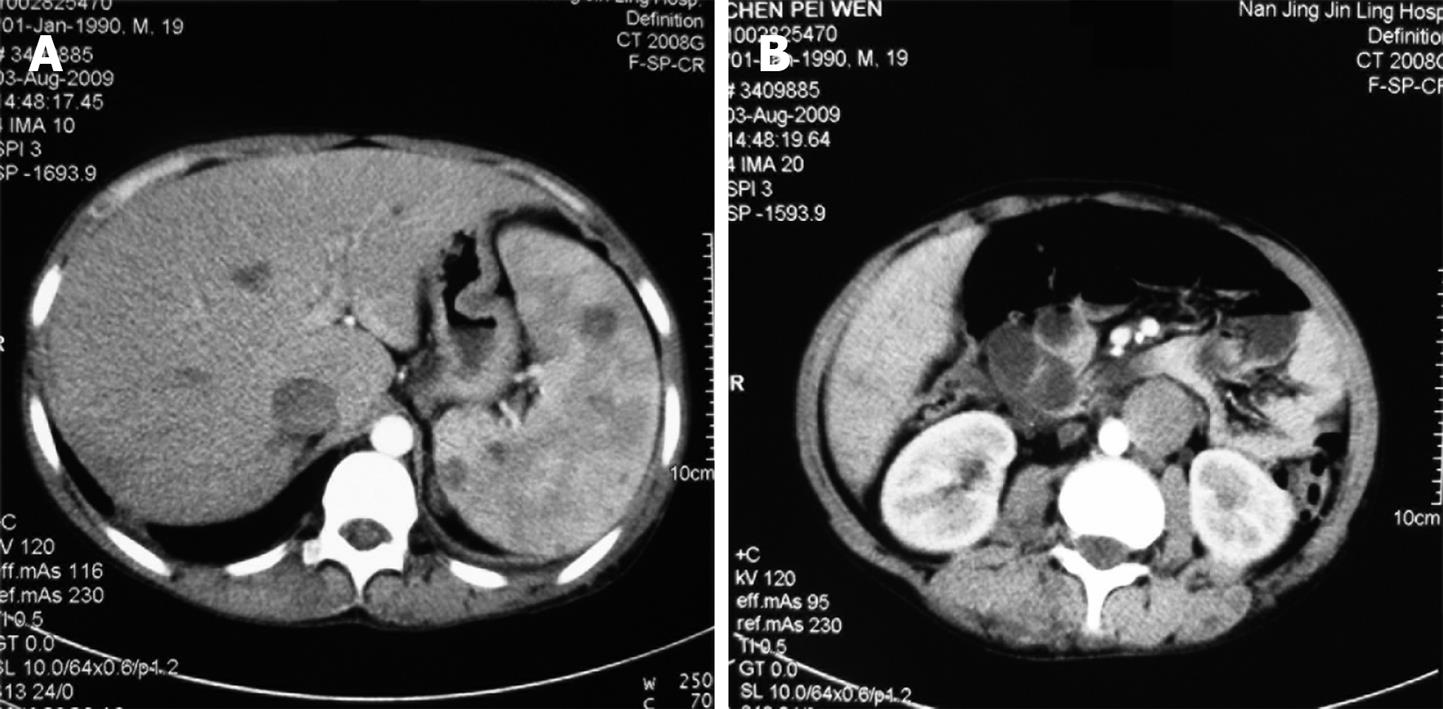
Figure 4 Contrast-enhanced abdominal computed tomography (CT) showing splenic hemangiomas (A) and left inferior vena cava (IVC) (B).
- Citation: Wang ZK, Wang FY, Zhu RM, Liu J. Klippel-Trenaunay syndrome with gastrointestinal bleeding, splenic hemangiomas and left inferior vena cava. World J Gastroenterol 2010; 16(12): 1548-1552
- URL: https://www.wjgnet.com/1007-9327/full/v16/i12/1548.htm
- DOI: https://dx.doi.org/10.3748/wjg.v16.i12.1548


