Copyright
©2010 Baishideng.
World J Gastroenterol. Mar 28, 2010; 16(12): 1548-1552
Published online Mar 28, 2010. doi: 10.3748/wjg.v16.i12.1548
Published online Mar 28, 2010. doi: 10.3748/wjg.v16.i12.1548
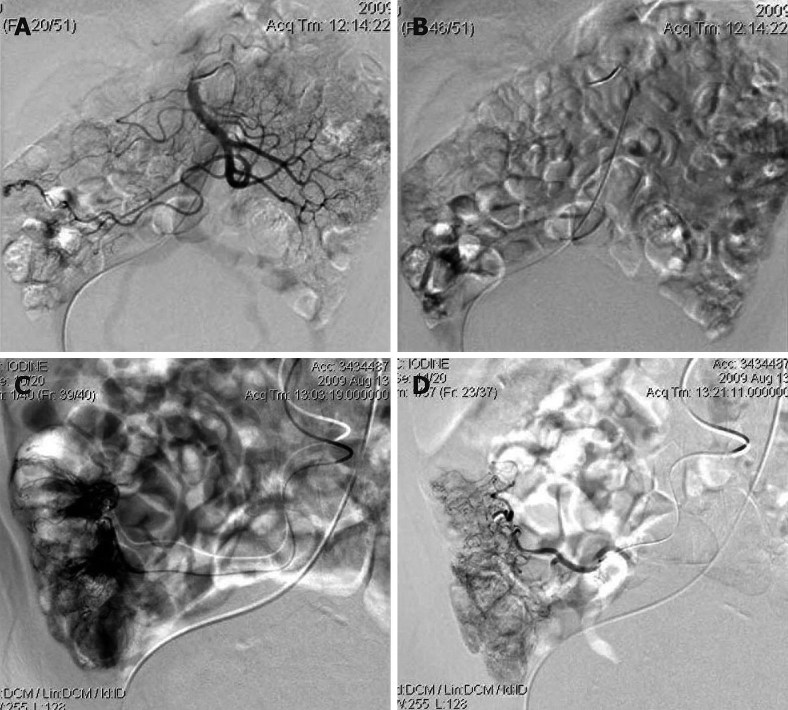
Figure 2 Angiograms demonstrate contrast opacification of the superior mesenteric artery and its branches.
A: Some abnormal small veins that were filled earlier in the arterial phase; B: Manipulus contrast extravasation into the terminal ileum; C: Abnormal ectatic slow-emptying veins and extravasation of contrast material after superselective catheterization; D: No active bleeding after superselective vessel embolism with gelfoam.
- Citation: Wang ZK, Wang FY, Zhu RM, Liu J. Klippel-Trenaunay syndrome with gastrointestinal bleeding, splenic hemangiomas and left inferior vena cava. World J Gastroenterol 2010; 16(12): 1548-1552
- URL: https://www.wjgnet.com/1007-9327/full/v16/i12/1548.htm
- DOI: https://dx.doi.org/10.3748/wjg.v16.i12.1548


