Copyright
©2010 Baishideng.
World J Gastroenterol. Mar 28, 2010; 16(12): 1545-1547
Published online Mar 28, 2010. doi: 10.3748/wjg.v16.i12.1545
Published online Mar 28, 2010. doi: 10.3748/wjg.v16.i12.1545
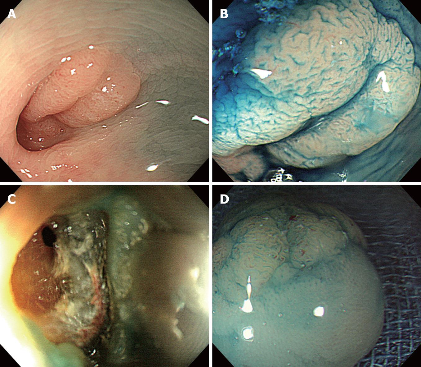
Figure 1 Colonoscopy and magnifying chromoendoscopy.
A: Colonoscopy showing a single diverticulum in the descending colon and a flat elevated polyp (15 mm in diameter) within the diverticulum; B: Magnifying chromoendoscopy using 0.4% indigo-carmine dye spraying revealing a type IV pit pattern according to Kudo’s classification; C: Endoscopy showing a pin-hole perforation immediately after endoscopic mucosal resection (EMR); D: Magnifying chromoendoscopy displaying negative neoplastic changes in the removed lesion.
- Citation: Fu KI, Hamahata Y, Tsujinaka Y. Early colon cancer within a diverticulum treated by magnifying chromoendoscopy and laparoscopy. World J Gastroenterol 2010; 16(12): 1545-1547
- URL: https://www.wjgnet.com/1007-9327/full/v16/i12/1545.htm
- DOI: https://dx.doi.org/10.3748/wjg.v16.i12.1545


