Copyright
©2010 Baishideng.
World J Gastroenterol. Mar 21, 2010; 16(11): 1418-1421
Published online Mar 21, 2010. doi: 10.3748/wjg.v16.i11.1418
Published online Mar 21, 2010. doi: 10.3748/wjg.v16.i11.1418
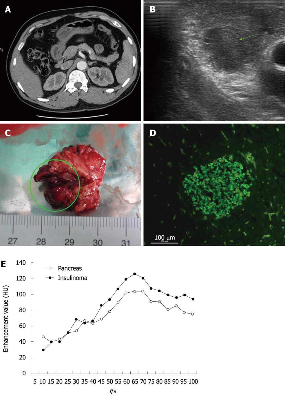
Figure 1 Clinical images related to this case and linear analysis of pancreatic dynamic enhanced spiral CT.
A: Negative result of abdominal enhanced spiral CT; B: We used intraoperative ultrasonography to detect the tumor and to determine the location relationship between the tumor and the main pancreatic duct. Fortunately, the tumor (green arrow) did not adjoin the main pancreatic duct. Therefore, we performed simple tumor resection; C: The insulinoma (green circle) (1.4 cm × 1.4 cm × 1.2 cm) was resected; D: Microscopic image of the insulinoma after immunostaining (× 400); E: Enhancement value of the pancreas and insulinoma at each time point after the onset of contrast material injection. The enhancement value peak of the tumor appeared at 65 s. As the interval of tumor-to-pancreas contrast was not obvious (mostly it is < 20 HU), the insulinoma was occult and difficult to find.
- Citation: Bao ZK, Huang XY, Zhao JG, Zheng Q, Wang XF, Wang HC. A case of occult insulinoma localized by pancreatic dynamic enhanced spiral CT. World J Gastroenterol 2010; 16(11): 1418-1421
- URL: https://www.wjgnet.com/1007-9327/full/v16/i11/1418.htm
- DOI: https://dx.doi.org/10.3748/wjg.v16.i11.1418


