Copyright
©2010 Baishideng.
World J Gastroenterol. Mar 14, 2010; 16(10): 1201-1208
Published online Mar 14, 2010. doi: 10.3748/wjg.v16.i10.1201
Published online Mar 14, 2010. doi: 10.3748/wjg.v16.i10.1201
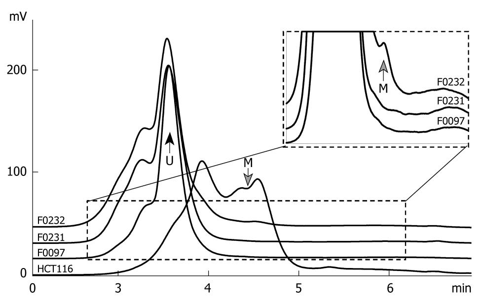
Figure 3 Chromatogram of the methylated and unmethylated GATA-4 CpG islands by denatured high performance liquid chromatography (DHPLC).
The 385-bp PCR product of the methylated and unmethylated GATA-4 were separated by DHPLC at a partial denaturing temperature 57.1°C and detected by a fluorescence (FL)-detector. The proportion of methylated GATA-4 in the tested samples was calculated according to the ratio of the peak area for the methylated GATA-4 to the total peak area for both the methylated and unmethylated GATA-4. The gray arrow points to the peak for the methylated GATA-4 (M) at the retention time 4.5 min; The black arrow points to the peak for the unmethylated GATA-4 (U) at the retention time 3.6 min. Genomic DNA of HCT116 was used as the GATA-4 methylation positive control. The inserted chart represents the dashed line surrounding the area. GATA-4 methylation was detected in the tested sample F0232.
-
Citation: Wen XZ, Akiyama Y, Pan KF, Liu ZJ, Lu ZM, Zhou J, Gu LK, Dong CX, Zhu BD, Ji JF, You WC, Deng DJ. Methylation of
GATA-4 andGATA-5 and development of sporadic gastric carcinomas. World J Gastroenterol 2010; 16(10): 1201-1208 - URL: https://www.wjgnet.com/1007-9327/full/v16/i10/1201.htm
- DOI: https://dx.doi.org/10.3748/wjg.v16.i10.1201


