Copyright
©2009 The WJG Press and Baishideng.
World J Gastroenterol. Mar 7, 2009; 15(9): 1057-1064
Published online Mar 7, 2009. doi: 10.3748/wjg.15.1057
Published online Mar 7, 2009. doi: 10.3748/wjg.15.1057
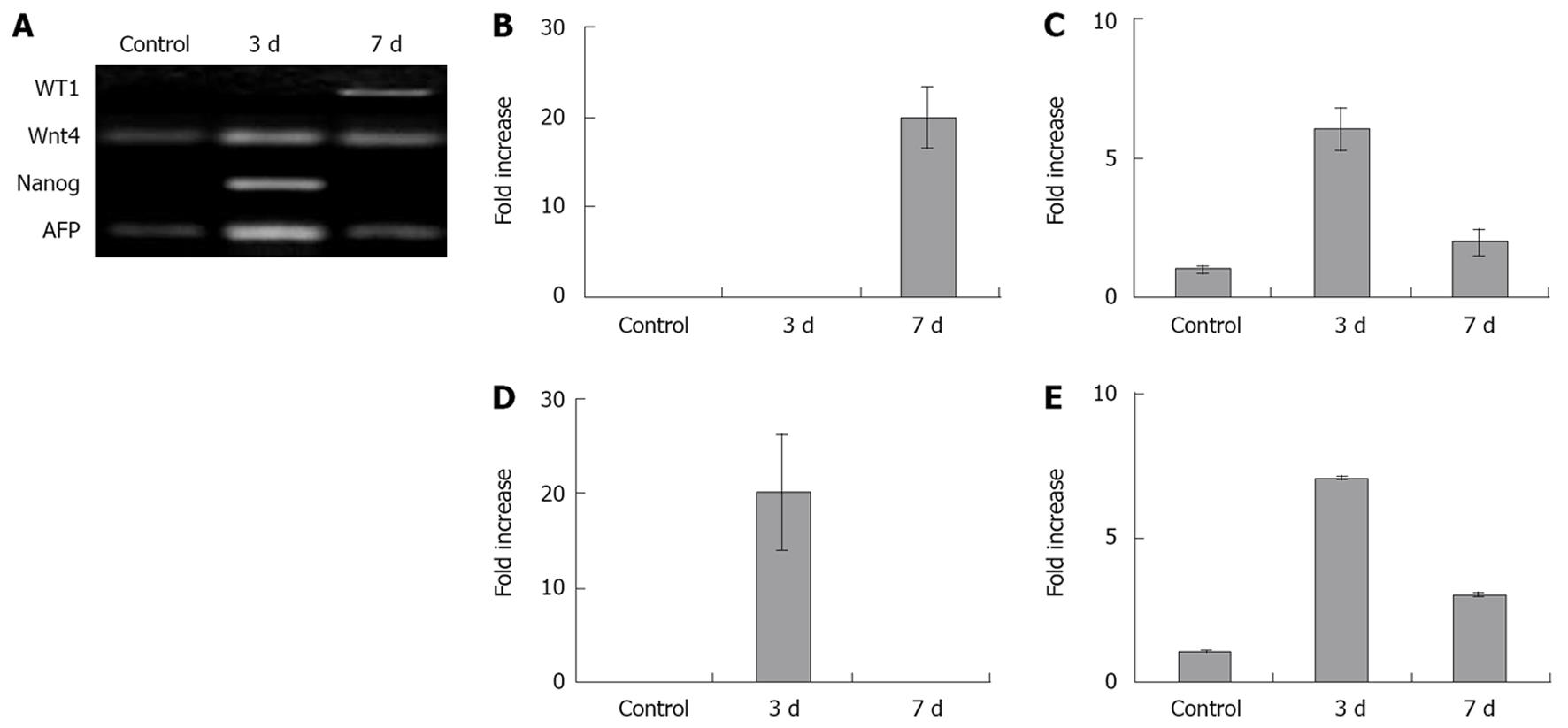
Figure 6 Activation of developmental genes in the regenerated liver at the wound site at days 3 and 7 after injury and fusion of the activated omentum.
The regenerating part of the liver (part of liver attached to the omentum) showed high expression levels (7 to 20-fold) of WT-1, Wnt-4, Nanog, AFP by RT-PCR (A) compared with normal rat adult liver tissue (control). Wnt-4 (C), Nanog (D), and AFP (E) were maximally activated at day 3, while WT-1 (B) showed maximal activation at day 7. Tissue from regenerated liver, from sites further away from the wound area (0.5 cm, 1.0 cm further away in the same lobe and from an uninjured lobe), showed reduced activation of WT-1, Wnt-4 and AFP genes (although higher in all cases compared with normal adult liver), suggesting that the regeneration stimulus ‘rippled’ throughout the liver from the wound area (data not shown). n = 3 in each bar and the differences amongst the bars within each of the figures B, C, D, and E are statistically significant (P < 0.05).
- Citation: Singh AK, Pancholi N, Patel J, Litbarg NO, Gudehithlu KP, Sethupathi P, Kraus M, Dunea G, Arruda JA. Omentum facilitates liver regeneration. World J Gastroenterol 2009; 15(9): 1057-1064
- URL: https://www.wjgnet.com/1007-9327/full/v15/i9/1057.htm
- DOI: https://dx.doi.org/10.3748/wjg.15.1057


