Copyright
©2009 The WJG Press and Baishideng.
World J Gastroenterol. Mar 7, 2009; 15(9): 1057-1064
Published online Mar 7, 2009. doi: 10.3748/wjg.15.1057
Published online Mar 7, 2009. doi: 10.3748/wjg.15.1057
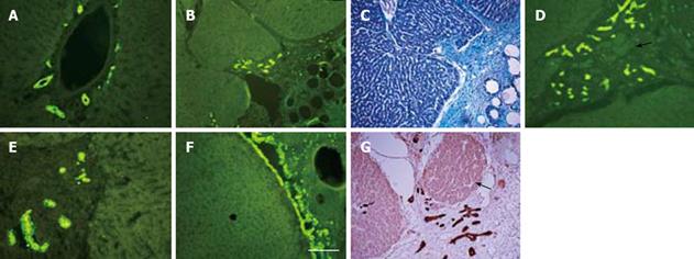
Figure 4 Immunostaining of normal and regenerated rat liver (from activated omentum) for cytokeratin-19, a marker of oval cells.
A: Normal or uninjured liver lobe showing widespread presence of oval cells in the lining of bile ducts lying around a central vein; B, D, E, G: Different areas of injured liver showing extensions of cytokeratin-19 positive bile ducts in the interlying tissue between the liver and the activated omentum; C: Tissue section shown in B stained with Trichrome to show the bile ducts lying in the interlying omental tissue; F: Occasionally, the growing edge of the liver lying in the interlying tissue was seen to be entirely covered with cytokeratin-19 positive cells; G: Islands of liver tissue, probably newly formed, were seen in the interlying tissue (white arrows; also seen in D); A, B, D-F were stained by immunofluorescence (green); G was stained by immunoperoxidase (brown). The horizontal white bar in F represents 100 &mgr;m for all pictures.
- Citation: Singh AK, Pancholi N, Patel J, Litbarg NO, Gudehithlu KP, Sethupathi P, Kraus M, Dunea G, Arruda JA. Omentum facilitates liver regeneration. World J Gastroenterol 2009; 15(9): 1057-1064
- URL: https://www.wjgnet.com/1007-9327/full/v15/i9/1057.htm
- DOI: https://dx.doi.org/10.3748/wjg.15.1057


