Copyright
©2009 The WJG Press and Baishideng.
World J Gastroenterol. Feb 21, 2009; 15(7): 829-835
Published online Feb 21, 2009. doi: 10.3748/wjg.15.829
Published online Feb 21, 2009. doi: 10.3748/wjg.15.829
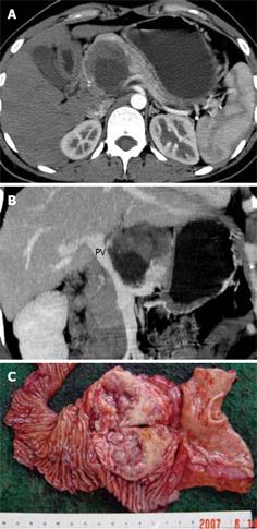
Figure 3 Pathologically confirmed benign SPT in a 30-year-old woman with abdominal pain for about 3 mo.
A: A heterogeneously low-attenuation mass was identified in the pancreatic head on the axial MDCT image at the arterial phase. The capsule was irregular and not attached to the adjacent, pancreas implying invasion outside the mass. The peripheral portion of the mass close to the duodenum was enhanced significantly and the duodenum was infiltrated; B: MPVR image revealed the irregularity and narrowing of the portal vein (PV) indicating vascular invasion; C: Invasion into the duodenum was revealed in the gross specimen of the tumor.
- Citation: Wang DB, Wang QB, Chai WM, Chen KM, Deng XX. Imaging features of solid pseudopapillary tumor of the pancreas on multi-detector row computed tomography. World J Gastroenterol 2009; 15(7): 829-835
- URL: https://www.wjgnet.com/1007-9327/full/v15/i7/829.htm
- DOI: https://dx.doi.org/10.3748/wjg.15.829


