Copyright
©2009 The WJG Press and Baishideng.
World J Gastroenterol. Feb 21, 2009; 15(7): 829-835
Published online Feb 21, 2009. doi: 10.3748/wjg.15.829
Published online Feb 21, 2009. doi: 10.3748/wjg.15.829
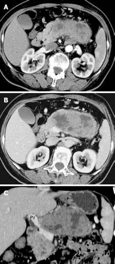
Figure 2 Malignant SPT in a 35-year-old woman with abdominal pain and mass for 6 mo.
A: A mass measuring 13 x 5.5 cm was detected on axial MDCT imaging in the pancreatic body. The mass was markedly enhanced and superimposed by abnormal enhancement of tiny vessels at the arterial phase. The interface between the tumor and the adjacent pancreatic parenchyma was blurred in that the tumor apparently infiltrated the surrounding pancreas; B: At the portal venous phase, the axial image demonstrated the heterogeneity of cystic and solid components inside the tumor. The peritumoral capsule was not smooth, which was consistent with capsular invasion. The portal vein was deformed by tumoral invasion; C: Coronal MPVR image showed the tumoral invasion resulting in the narrowing and irregularity of portal vein (PV).
- Citation: Wang DB, Wang QB, Chai WM, Chen KM, Deng XX. Imaging features of solid pseudopapillary tumor of the pancreas on multi-detector row computed tomography. World J Gastroenterol 2009; 15(7): 829-835
- URL: https://www.wjgnet.com/1007-9327/full/v15/i7/829.htm
- DOI: https://dx.doi.org/10.3748/wjg.15.829


